
All iLive content is medically reviewed or fact checked to ensure as much factual accuracy as possible.
We have strict sourcing guidelines and only link to reputable media sites, academic research institutions and, whenever possible, medically peer reviewed studies. Note that the numbers in parentheses ([1], [2], etc.) are clickable links to these studies.
If you feel that any of our content is inaccurate, out-of-date, or otherwise questionable, please select it and press Ctrl + Enter.
Treatment of osteoarthritis: use of glucocorticosteroids
Medical expert of the article

Last reviewed: 04.07.2025
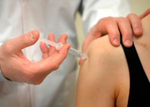 ">
">Systemic use of corticosteroids in osteoarthritis is not indicated, but intra-articular and periarticular injections of prolonged (depot) forms of corticosteroids provide a significant, albeit temporary, symptomatic effect.
The variety of NSAIDs on the modern pharmaceutical market and the abundance of often contradictory information about their pharmacodynamics, efficacy and safety make it difficult to choose a drug. It is not always possible to extrapolate the results of a multicenter controlled study of efficacy to a specific patient. As noted above, the main feature by which NSAIDs differ from each other is their tolerability.
There is no evidence of the superiority of some NSAIDs over others in terms of analgesic and anti-inflammatory properties. In addition, in light of recent discoveries of more complex mechanisms of COX-1 and COX-2 participation in pathological and physiological processes, it is becoming obvious that selective and even specific (coxibs) COX-2 inhibitors are not “ideal” NSAIDs. In order to ensure effective and safe treatment, first of all, a thorough examination of the patient is necessary to exclude risk factors for the development of side effects. If a risk of gastropathy is detected, it is rational to prescribe selective or specific COX-2 inhibitors. If a non-selective NSAID shows significant efficacy in a particular patient, it can be prescribed in combination with misoprostol, proton pump inhibitors or H2 - receptor antagonists.
In the presence of signs of renal failure, it is inappropriate to prescribe NSAIDs, however, if the appointment of NSAIDs is necessary, preference should be given to specific COX-2 inhibitors, and treatment should be carried out under careful monitoring of the level of creatinine in the blood serum. Patients at risk of thrombosis during treatment with COX-2 inhibitors should continue taking acetylsalicylic acid in low doses and carefully monitor the state of the gastrointestinal tract.
When choosing NSAIDs from the non-selective group for an elderly patient, preference should be given to propionic acid derivatives, which are short-lived NSAIDs (rapidly absorbed and eliminated), which do not accumulate when metabolic processes are disrupted. If the patient does not belong to the risk group for developing side effects, treatment can be started with either a non-selective or a selective or specific COX-2 inhibitor. If the drug is ineffective or insufficiently effective, it must be changed.
Main drugs of depot forms of corticosteroids
Preparation |
Content of active substance in 1 ml of suspension |
Kenalog 40 |
40 mg triamcinolone acentonide |
Diprospan |
2 mg betamethasone disodium phosphate and 5 mg betamethasone dipropionate |
Depo-medrol |
40 mg methylprednisolone acetate |
A feature of corticosteroid preparations used for intra-articular administration is a prolonged anti-inflammatory and analgesic effect. Taking into account the duration of the effect, depot corticosteroids can be arranged in the following order:
- hydrocortisone acetate - is available in the form of a microcrystalline suspension in 5 ml vials (125 mg of the drug); when administered intra-articularly, it is practically not absorbed from the cavity, the effect lasts from 3 to 7 days; due to the relatively weak and short effect, it has been used extremely rarely lately;
- triamcinolone acetonide - is available in the form of an aqueous crystalline suspension, in ampoules of 1 and 5 ml (40 mg/ml); the anti-inflammatory and analgesic effect occurs 1-2 days after injection and lasts 2-3 (less often 4) weeks; the main disadvantage is the frequent development of atrophy of the skin and subcutaneous fat, necrosis of tendons, ligaments or muscles at the injection site;
- methylprednisolone acetate - is available in the form of an aqueous suspension, in ampoules of 1, 2 and 5 ml (40 mg/ml); in terms of duration and severity of effect, it is almost no different from the drug triamcinolone acetonide; when used in recommended doses, the risk of developing atrophy and necrosis of soft tissues at the injection site is minimal; has virtually no mineralocorticoid activity;
- a combination drug (trade names registered in Ukraine - Diprospan, Flosteron), containing 2 mg of betamethasone disodium phosphate (highly soluble, rapidly absorbed ester, provides a rapid effect) and 5 mg of betamethasone dipropionate (poorly soluble, slowly absorbed depot fraction, has a prolonged effect), available in 1 ml ampoules, the composition of the drug determines a rapid (already 2-3 hours after intra-articular administration) and prolonged (3-4 weeks) effect; the micronized structure of the suspension crystals ensures painless injections.
Local intra-articular injection of triampinolone hexacetonide caused a short-term reduction in pain in knee joints affected by osteoarthrosis; treatment results were better in cases of preliminary aspiration of exudate from the joint cavity before injection. R.A. Dieppe et al. (1980) demonstrated that local intra-articular injection of corticosteroids leads to a more pronounced reduction in pain than placebo.
The main indications for the use of corticosteroids in osteoarthrosis are persistent synovitis despite conservative treatment, as well as persistent inflammation of the periarticular tissues (tendovaginitis, bursitis, etc.). When planning intra-articular administration of prolonged glucocorticosteroids, it is necessary to remember that drugs of this group are contraindicated in infectious arthritis of various etiologies, infection of the skin and subcutaneous fat or muscles in the injection area, sepsis, hemarthrosis (hemophilia, trauma, etc.), intra-articular fractures. In case of persistent pain syndrome and the absence of synovitis that is not relieved by conservative therapy, glucocorticosteroids should not be injected into the joint, but should be administered periarticularly. At stages III-IV according to Kellgren and Lawrence, intra-articular injections of glucocorticosteroids should be used with extreme caution, only if conservative measures are ineffective.
An important requirement when performing intra-articular injections is compliance with aseptic rules:
- the doctor's hands must be clean, preferably wearing surgical gloves,
- Only disposable syringes are used,
- after drawing the drug into the syringe, immediately before administration, the needle is changed to a sterile one,
- evacuation of intra-articular fluid and administration of the drug must be done with different syringes,
- the injection area is treated with a 5% alcohol solution of iodine, then with 70% alcohol,
- After administration, the injection site is pressed with a cotton swab soaked in 70% alcohol and fixed with a plaster or bandage for at least 2 hours,
- During the procedure, the staff and the patient should not talk.
After inserting the needle into the joint cavity, it is necessary to aspirate the maximum amount of synovial fluid, which already contributes to some analgesic effect (intra-articular pressure decreases, mechanical and biochemical inducers of inflammation are removed from the cavity with the synovial fluid), and also frees up space for the subsequent administration of the drug.
According to HJ Kreder et al. (1994), the negative effect of intra-articular glucocorticosteroid injections in rabbits was potentiated by their motor activity. After intra-articular administration of depot forms of glucocorticosteroids, it is advisable not to load the joint for some time, since observing a period of rest after the injection contributes to a more pronounced and prolonged effect.
Since animal studies have demonstrated the ability of glucocorticosteroids to damage articular cartilage, and frequent intra-articular injections of depot forms of glucocorticosteroids are associated with the destruction of intra-articular tissues, injections are not recommended to be administered more often than 3-4 times a year. However, H.W. Balch et al. (1977), who retrospectively evaluated joint radiographs after repeated injections over a period of 4-15 years, argued that rational use of repeated injections of these drugs does not lead to an acceleration of disease progression according to radiographic data.
Complications of local glucocorticosteroid therapy can be divided into intra-articular and extra-articular:
Intra-articular:
- Ineffectiveness of intra-articular GCS therapy due to resistance of joint tissues to glucocorticosteroids is observed in 1-10% of patients. It is believed that the mechanism of this process is based on a deficiency of GK receptors in the inflamed synovial tissue,
- increased pain and swelling of the joint is observed in 2-3% of patients, which is associated with the development of phagocytosis of hydrocortisone crystals by leukocytes of the synovial fluid;
- osteoporosis and osteochondral destruction. JL Hollander, analyzing the results of long-term treatment of 200 patients, along with a good clinical effect, observed rapid progression of osteoporosis in 16% of patients, erosion of articular cartilage in 4% and an increase in bone destruction of articular surfaces in 3% of patients,
- hemarthrosis; G.P. Matveenkov and co-authors (1989) observed two cases of hemarthrosis during 19,000 joint punctures;
- infection of the joint cavity with subsequent development of purulent arthritis; most often, infection occurs in the knee joint, as a rule, signs of inflammation appear 3 days after the injection.
Extra-articular:
- skin atrophy at the injection site occurs when the drug enters extra-articular tissues and is observed mainly after injections of glucocorticosteroids into small joints: jaw, interphalangeal, metacarpophalangeal; skin atrophy has been described after injections into the knee joint;
- linear hypopigmentation extending proximally from the joint;
- periarticular calcification - may be accompanied by atrophy of the skin over the joints,
- tissue granulomatous reactions,
- ligament and tendon ruptures, pathological bone fractures.