
All iLive content is medically reviewed or fact checked to ensure as much factual accuracy as possible.
We have strict sourcing guidelines and only link to reputable media sites, academic research institutions and, whenever possible, medically peer reviewed studies. Note that the numbers in parentheses ([1], [2], etc.) are clickable links to these studies.
If you feel that any of our content is inaccurate, out-of-date, or otherwise questionable, please select it and press Ctrl + Enter.
Submandibular salivary gland
Medical expert of the article

Last reviewed: 07.07.2025
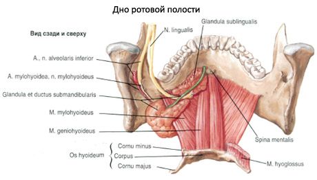
The submandibular salivary gland (glandula submandibularis) is paired, of mixed secretion type, and has a thin capsule. It is located in the area of the triangle of the neck of the same name. The superficial plate of the cervical fascia and skin are adjacent to the gland on the outside. The inner surface of the gland is in contact with the hyoglossus and styloglossus muscles. From above, the gland reaches the inner surface of the body of the lower jaw.
The upper edge of the gland adjoins the lower jaw, and the upper surface - the mylohyoid muscle. Having rounded the posterior edge of the specified muscle, the submandibular salivary gland is located on its upper surface and comes into contact with the postero-external surface of the sublingual SG. The posterior edge of the submandibular salivary gland reaches the capsule of the parotid SG and the medial pterygoid muscle. The excretory duct begins from the upper inner edge of the gland, then penetrates into the gap between the mylohyoid and hyoglossus muscles. Along the inner surface of the sublingual SG, the excretory duct goes forward and upward and opens in the anterior part of the floor of the oral cavity on the sublingual papilla.
The submandibular salivary gland is surrounded on all sides by a capsule formed by the superficial plate of the cervical fascia. The latter, splitting, forms a sheath for the submandibular salivary gland, the outer plate of which is attached to the lower edge of the lower jaw, the inner - to the line of attachment of the mylohyoid muscle. Between the submandibular salivary gland and the sheath there is a layer of loose cellular tissue. The submandibular space is limited below by the superficial leaflet of the proper fascia of the neck, above - by the fascial case of the mylohyoid muscle, loose fascia covering the mylohyoid muscle, and the superior constrictor of the pharynx. From the submandibular space, the pathological process spreads to the anterior part of the parapharyngeal space and the sublingual cellular space. The spread into the parotid cellular space is prevented by a strong aponeurosis, which goes from the case of the sternocleidomastoid muscle to the angle of the lower jaw. The facial artery, the anterior facial vein, and the lymph nodes are also located in this closed space. The latter collect lymph from the upper and lower lips, oral cavity, tongue, lower jaw, and pharynx.
The anterior part of the gland is in contact with the posterior edge of the mylohyoid muscle. The lateral surface of the gland is adjacent to the facial artery and vein, and is also adjacent to the lymph nodes of the same name. Wharton's duct of the gland (ductus submandibularis) is directed forward, is adjacent to the sublingual salivary gland and opens with an orifice on the sublingual papilla, next to the frenulum of the tongue.
Innervation: secretory (parasympathetic) - fibers of the facial nerve - from the chorda tympani and submandibular ganglion, sympathetic - from the external carotid plexus.
In the submandibular sheath, slightly above (2-8 mm) the posterior belly of the digastric muscle, the hypoglossal nerve (XII pair of cranial nerves) passes, which is accompanied by the lingual vein.
The sensory lingual nerve passes through the upper part of the submandibular triangle.
The submandibular salivary gland is innervated by the chorda tympani (from the anterior part of the floor of the oral cavity, along the facial nerve) through the submandibular ganglion and the sympathetic nerves accompanying the facial artery. Lymph drainage occurs to the lymph nodes at the lower pole of the parotid salivary gland and to the deep jugular lymph nodes.
Blood supply: glandular branches of the facial artery. Venous drainage: submandibular vein.
The facial artery, being a branch of the external carotid artery, passes into the submandibular triangle from under the posterior belly of the digastric muscle and the stylohyoid muscle and penetrates the submandibular gland at its posterior edge. At the level of the anterior edge of the masseter muscle, the facial artery exits the gland onto the face, bending over the edge of the lower jaw (its pulsation is easily palpated here).
The submandibular salivary gland is supplied with blood by branches of the facial, lingual and mental arteries. The venous network in this area is formed by the anterior facial and retromandibular veins, which flow into the common facial vein. The anterior facial vein accompanies the facial artery, is located at the lower edge of the lower jaw behind the artery, penetrates the capsule of the gland and runs along its anterior surface.
Lymph drainage: to the submandibular lymph nodes.
What do need to examine?