
All iLive content is medically reviewed or fact checked to ensure as much factual accuracy as possible.
We have strict sourcing guidelines and only link to reputable media sites, academic research institutions and, whenever possible, medically peer reviewed studies. Note that the numbers in parentheses ([1], [2], etc.) are clickable links to these studies.
If you feel that any of our content is inaccurate, out-of-date, or otherwise questionable, please select it and press Ctrl + Enter.
A newborn's skull
Medical expert of the article

Last reviewed: 06.07.2025
The skull of a newborn has a number of significant features. The cranial skull, as a result of active brain growth and early formation of the sense organs, is 8 times larger in volume than the facial skull. In an adult, due to the complete development of the masticatory apparatus, the cranial skull is only 2 times larger than the facial skull. The eye sockets of a newborn are wide. The base of the skull, compared to the vault, lags behind in growth, the bones are connected to each other by means of wide cartilaginous and connective tissue layers. The tubercles of the frontal and parietal bones are well defined, and therefore, when examining the skull from above, it appears quadrangular. The frontal bone consists of two halves, the superciliary arches are absent, and the frontal sinus is not yet present. The jaws are underdeveloped, which causes the small height of the facial skull. The lower jaw consists of two parts (two halves). The parts of the temporal bone are separated from each other by well-defined connective tissue or cartilaginous layers, the mastoid process is not developed. On the bones of the skull, the muscular tubercles and lines are not expressed.
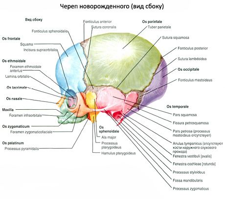
The most characteristic feature of the newborn's skull is the fontanelles (fonticuli). They are non-ossified connective tissue (membranous) areas of the cranial vault. There are six fontanelles in total: two lie along the midline of the cranial vault and four lateral fontanelles
- The largest is the anterior (frontal) fontanelle (fonticulus anterior). It is diamond-shaped, located between the two parts of the frontal bone and both parietal bones, and closes in the 2nd year of life.
- The posterior (occipital) fontanelle (fonticulus posterior) is triangular in shape. It is located between the two parietal bones in front and the occipital squama at the back; it closes in the 2nd month of life.
- The lateral fontanelles are paired, two on each side.
- The anterior sphenoid fontanelle (fonticulus sphenoidalis) is located at the junction of the large wing of the sphenoid bone with the frontal, parietal bones and squama of the temporal bone; it closes in the 2nd-3rd month of life.
- The posterior - mammillary fontanelle (fonticulus mastoideus) - is formed by the temporal, parietal bones and occipital squama; it overgrows in the 2nd-3rd month of life.
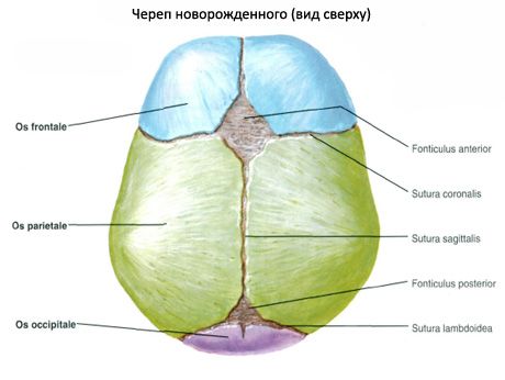
The sutures between the bones of the cranial vault are not formed, the edges of the bones are even. Only in the 3rd year of a child's life, the bones of the skull begin to develop teeth, which gradually increase in size and enter the spaces between the teeth of the neighboring bone. This is how the serrated sutures are formed. From the description of the skull of a newborn, it is clear that by the time of birth its development is far from complete. It continues in the following years of life.
 [
[