
All iLive content is medically reviewed or fact checked to ensure as much factual accuracy as possible.
We have strict sourcing guidelines and only link to reputable media sites, academic research institutions and, whenever possible, medically peer reviewed studies. Note that the numbers in parentheses ([1], [2], etc.) are clickable links to these studies.
If you feel that any of our content is inaccurate, out-of-date, or otherwise questionable, please select it and press Ctrl + Enter.
Sarcoidosis and glaucoma
Medical expert of the article

Last reviewed: 04.07.2025
Sarcoidosis is a systemic disease characterized by the formation of noncaseating, granulomatous inflammatory infiltrates in the lungs, skin, liver, spleen, central nervous system, and eyes.
Ocular involvement occurs in 10-38% of patients with systemic sarcoidosis. Ocular sarcoidosis, manifested as anterior, middle, posterior, or panuveitis, leads to the development of chronic granulomatous uveitis.
Epidemiology of sarcoidosis-associated glaucoma
Sarcoidosis is 8-10 times more common in African Americans than in whites, with an incidence of 82 cases per 100,000. The disease can develop at any age, but is most common in patients aged 20-50 years. About 5% of uveitis cases in adults and 1% of uveitis cases in children are associated with sarcoidosis. Seventy percent of cases of sarcoidosis involve the anterior segment, with posterior segment involvement occurring in less than 33%. Approximately 11-25% of patients with sarcoidosis develop secondary glaucoma, most often with anterior segment involvement. African American patients with sarcoidosis are more likely to develop secondary glaucoma and blindness.
What causes sarcoidosis?
The development of ocular hypertension and glaucoma in patients with sarcoidosis occurs with obstruction of the trabecular meshwork as a result of the chronic inflammatory process, as well as with closure of the anterior chamber angle due to the formation of peripheral anterior and posterior synechiae and iris bombage. Neovascularization of the anterior segment of the eye and prolonged use of glucocorticoids can also lead to impaired outflow of intraocular fluid.
Symptoms of glaucoma associated with sarcoidosis
Most adults with sarcoidosis have lung involvement, cough, shortness of breath, wheezing, or shortness of breath with exertion. Other manifestations of sarcoidosis include systemic symptoms such as fever, fatigue, and weight loss. Often, there may be no symptoms at diagnosis. When the eyes are involved, patients typically complain of eye pain, redness, sensitivity to light, floaters, blurred vision, or decreased visual acuity.
Course of the disease
Sarcoidosis of the eye can be acute and self-limited or have a chronic recurrent or continuous course. The prognosis for chronic sarcoidosis uveitis is the most unfavorable due to the development of complications (glaucoma, cataracts or macular edema).
Diagnosis of sarcoidosis-associated glaucoma
Differential diagnosis of sarcoidosis should include other conditions that cause granulomatous panuveitis, such as Vogt-Koyanagi-Harada syndrome, sympathetic ophthalmia, and tuberculosis. Syphilis, Lyme disease, primary intraocular lymphoma, and pars planitis should be considered as ocular involvement.
 [ 9 ]
[ 9 ]
Laboratory research
The diagnosis of sarcoidosis is made when noncaseating or nonnecrotic granulomas or granulomatous inflammation are detected in a tissue biopsy of a patient in whom other granulomatous diseases (tuberculosis and fungal infections) have been excluded. When sarcoidosis is initially diagnosed, chest radiography and serum angiotensin-converting enzyme (ACE) levels should be measured. Serum lysozyme levels may be elevated, which is less specific than ACE levels, a marker of the disease. However, ACE levels may be elevated in healthy children, so this criterion is of less diagnostic value in pediatric patients. Increased ACE levels have been shown in the intraocular and cerebrospinal fluid in patients with sarcoidosis of the eye and central nervous system (sarcoidosis uveitis and neurosarcoidosis, respectively). Additional studies that help confirm the diagnosis include immunological tolerance testing, pulmonary function tests, Ga-enhanced testing, chest computed tomography, bronchoalveolar lavage, and transbronchial biopsy.
Ophthalmological examination
Eye involvement in sarcoidosis is usually bilateral, although it may be unilateral or with marked asymmetry. Granulomatous uveitis most often develops in sarcoidosis, but nongranulomatous uveitis may also develop. Examination reveals granulomas of the skin and orbit, enlarged lacrimal glands, and nodular formations of the conjunctiva of the eyelids and cheeks. Examination of the cornea usually reveals large sebaceous precipitates and coin-shaped infiltrates; less commonly, endothelial opacity is observed in the lower part of the cornea. With extensive posterior and peripheral anterior synechiae, intraocular pressure increases and secondary inflammatory glaucoma develops, associated with closure of the angle of the anterior chamber or bombage of the iris. Often, with severe inflammation of the anterior segment of the eye, Koeppe and Busacca nodules are detected on the iris.
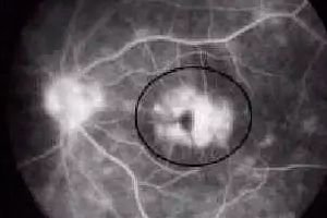
Posterior segment involvement in sarcoidosis is less common than anterior segment involvement. Examination of the vitreous often reveals inflammation with opacities and accumulation of inflammatory products in its inferior portion. Fundus examination may reveal various changes, including peripheral retinal vasculitis, peripheral snowdrift-type exudation, hemorrhages, retinal exudates, perivascular nodular granulomatous lesions, Dalen-Fuchs nodules, retinal and subretinal neovascularization, and neovascularization of the optic disc. Granulomas may also be found in the retina, choroid, or optic nerve. Decreased visual acuity in sarcoidosis occurs due to the formation of cystoid macular edema, optic neuritis with granulomatous infiltration, and secondary glaucoma.
Who to contact?
Treatment of glaucoma associated with sarcoidosis
The main method of treating both systemic and ocular sarcoidosis is glucocorticoid therapy. In case of damage to the anterior segment of the eye, they are used locally or orally. Systemic treatment is necessary for bilateral posterior uveitis. In sarcoidosis, other immunosuppressants have been shown to be effective, for example, cyclosporine and methotrexate. They should be used in case of chronic course of the disease and the need for long-term treatment with glucocorticoids. Treatment of glaucoma with drugs that reduce the formation of intraocular fluid should be carried out for as long as possible. Argon laser trabeculoplasty is often ineffective. The method of choice for pupillary block is laser iridotomy or surgical iridectomy. If the intraocular pressure remains high, either a filtering operation or implantation of a tubular drainage is recommended. The effectiveness of surgical treatment increases if the inflammatory process is stopped before the operation. Antimetabolites are recommended for trabeculectomy, especially in African American patients.