
All iLive content is medically reviewed or fact checked to ensure as much factual accuracy as possible.
We have strict sourcing guidelines and only link to reputable media sites, academic research institutions and, whenever possible, medically peer reviewed studies. Note that the numbers in parentheses ([1], [2], etc.) are clickable links to these studies.
If you feel that any of our content is inaccurate, out-of-date, or otherwise questionable, please select it and press Ctrl + Enter.
Pneumonitis in adults and children
Medical expert of the article

Last reviewed: 12.07.2025
Pulmonologists classify pneumonitis as an interstitial lung disease, the distinctive feature of which is damage to the tissues that support the intralobular air exchange part of the lungs and form its most important structures – the alveoli.
Epidemiology
The actual statistics of pneumonitis are unknown. According to some data, the prevalence of idiopathic interstitial pneumonia (which many define as idiopathic pneumonitis) per 100 thousand people in the European continent and North America is estimated at 7-50 cases with a tendency to constant growth. [ 1 ]
Chronic pneumonitis is observed in almost 5% of patients with this disease.
Acute lupus pneumonitis affects up to 10% of patients with SLE. And radiation pneumonitis after radiation therapy for advanced lung cancer is observed in three out of ten patients. [ 2 ]
According to the WHO, pneumonitis is one of the top three causes of death in older people due to respiratory failure. [ 3 ]
Causes pneumonitis
Due to the lack of terminological clarity, some doctors continue to interpret the name "pneumonitis" as a general designation of inflammatory processes in the lungs, but it should be immediately explained what the difference is between pneumonitis and pneumonia. First of all, these are etiological differences: if inflammation in pneumonia is caused by an infection - bacterial, viral or fungal, then the causes of pneumonitis are not associated with these infections, and the inflammation is immunologically mediated. Thus, viral pneumonitis as a diagnosis contradicts the pathogenetic essence of the disease identified by researchers, and publications on pneumonitis caused by viruses (RSV, Varicella Zoster, HSV or Cytomegalovirus) date back to the 70-90s of the last century.
It is also necessary to take into account the peculiarities of alteration of lung tissue: inflammation in cases of pneumonia has an exudative character with infiltration of the parenchyma, and pneumonitis is characterized by fibrous changes in the tissues of the alveolar and intralobular interstitium.
Depending on the etiology, there are different types or kinds of this lung disease, including pneumonitis in children, which develops for the same reasons.
Inflammation of the interstitium caused by an immune response to long-term inhaled airborne substances (aeroallergens) is defined as hypersensitivity pneumonitis or hypersensitivity pneumonitis; a simpler definition is allergic pneumonitis, which is often called exogenous allergic alveolitis. Triggers for the immune reaction leading to damage to the pulmonary interstitium may be dust containing animal or plant proteins (inhaled during agricultural and other work). This type includes the so-called "bird fancier's lung" - the result of an immune reaction to proteins in bird feathers and their dry droppings. [ 4 ]
If serologic testing of peripheral blood reveals elevated levels of eosinophils involved in hypersensitivity reactions, specialists may diagnose eosinophilic pneumonitis (also called Löffler syndrome or acute eosinophilic pneumonia ) or hypersensitivity reactive pneumonitis. When low-molecular-weight chemicals present in the air are inhaled, either as gases or as aqueous dispersions, chemical pneumonitis is diagnosed. And when lung damage is caused by inhaling toxic substances, toxic pneumonitis may develop. [ 5 ]
What is drug-induced pneumonitis, more details in the publication - Drug-induced lung lesions. For example, pneumonitis is one of the side effects of such antitumor drugs as Azathioprine, Nivolumab, Cyclophosphamide, Tocilizumab, Procarbazine, etc. In addition, foreign specialists highlight immune pneumonitis - a side effect of cancer immunotherapy using so-called immune checkpoint inhibitors: drugs Ipilimumab and Tremelimumab.
Aspiration pneumonitis, caused by the contents of the stomach entering the lower respiratory tract (Mendelson's syndrome or acid-aspiration pneumonitis caused by food, which often accompanies myasthenia gravis), is singled out separately, as well as that which occurs after nasogastric intubation or under general anesthesia, which causes vomiting. [ 6 ]
Obstructive pneumonitis is most often associated with airway obstruction by a tumor, such as in patients with squamous cell lung cancer.
As a result of exposure to ionizing radiation on the lungs - during radiation therapy of malignant neoplasms in the mediastinal region - radiation pneumonitis occurs; other definitions are post-radiation or radiation pneumonitis.
Desquamative or destructive pneumonitis – with disruption of the interstitial structure – can have any etiology, including long-term smoking. [ 7 ]
In patients with autoimmune diseases, non-specific pneumonitis is observed. Thus, in diffuse autoimmune disease of connective tissue – systemic lupus erythematosus – acute or chronic lupus pneumonitis or lupus pneumonitis is observed in almost half of the cases. [ 8 ]
Such a complication of the terminal stage of progressive renal failure as uremic pneumonitis is associated with diffuse disturbances in the permeability of alveolar capillary membranes, as well as interstitial and intraalveolar edema against the background of a decrease in blood clotting factors due to the high content of amino acid and protein metabolic products in it - urea nitrogen.
Quite often, the causes of pneumonitis cannot be determined, and then idiopathic pneumonitis is diagnosed, which may be called idiopathic fibrosing alveolitis.
Risk factors
The main risk factors for developing pneumonitis include:
- smoking;
- professions related to agriculture (harvesting grain crops, haymaking, poultry farming);
- tendency to allergic reactions;
- exposure to various substances in the air (in the workplace or in the environment);
- long-term use of certain medications;
- chemotherapy, immunotherapy and radiation therapy for cancer;
- the presence of systemic autoimmune diseases.
The risk of gastric aspiration into the lungs and the development of aspiration pneumonitis is increased in trauma, epileptic seizures, esophageal motility disorders, and severe gastroesophageal reflux. [ 9 ]
Pathogenesis
In pneumonitis, the pathogenesis of damage to the connective tissue interstitium, elastic walls of the alveoli and interalveolar septa is caused by a disruption of their structure at the cellular level and progressive fibrosis.
The interstitium consists of fibers (elastic and collagen), fibroblasts, connective tissue macrophages (histiocytes), neutrophils and some other cellular components.
The reaction of autoimmune antibodies to an antigen leads to increased division of effector T cells - T-helper lymphoid cells of the second type (Th2), which stimulate the cellular immune response to non-microbial foreign substances that are allergens.
The answer lies in the stimulation of proinflammatory cytokines, chemokines, NK and B lymphocytes of the alveolar interstitium tissues and an increase in the activity of transforming growth factor (TGF-β) and fibroblast growth factor (FGFR1-3). This causes an intensive proliferation of normal fibroblasts, as well as a multiple increase in the number of myofibroblasts (smooth muscle fibroblasts) present in the lung tissues, producing proteins and proteases of the extracellular matrix. [ 10 ]
Symptoms pneumonitis
Based on symptoms and instrumental diagnostic data, acute, subacute and chronic pneumonitis are classified.
Typically, the first signs of pneumonitis are dyspnea (shortness of breath) and a dry, hacking cough.
Pneumonitis can progress differently in different patients, but the most common symptoms include:
- difficulty breathing;
- discomfort in the mediastinal area;
- general weakness and increased fatigue;
- loss of appetite and unexplained weight loss;
- pulmonary hemorrhage.
In acute interstitial pneumonitis, cough may produce thick mucous sputum, and breathing difficulties in many cases progress rapidly, leading to severe respiratory failure at a later stage.
Bilateral or bilateral pneumonitis develops when the alveolar interstitium of both lungs is damaged.
In addition to shortness of breath and cough, symptoms of radiation pneumonitis include fever, heaviness and pain in the chest.
In lupus pneumonitis, there is a nonproductive cough with bleeding.
Pneumonitis in lung cancer is manifested by a prolonged cough with shortness of breath and hoarseness of the voice, as well as chest pain (especially severe with a deep breath). And with a certain localization of the primary tumor or its growth, obstructive pneumonitis in lung cancer can develop with a decrease in its volume - atelectasis of the lung, which leads to the development of respiratory distress syndrome. [ 11 ]
Complications and consequences
What is the danger of pneumonitis? If left untreated or if treatment is started late, pneumonitis can cause complications and consequences such as:
- irreversible damage to the pulmonary alveoli in the form of pulmonary fibrosis, as well as pneumosclerosis;
- pulmonary hypertension;
- right ventricular heart failure (pulmonary heart disease);
- respiratory failure, lung failure and death.
Diagnostics pneumonitis
Clinical diagnosis of pneumonitis involves taking a complete medical history and an extensive examination of the respiratory organs.
Necessary tests include a general and biochemical blood test; immunological blood test – for antigen-specific IgG antibodies and other circulating immune complexes in the blood.
A diagnostic bronchoalveolar lavage (washing) and laboratory testing of the resulting fluid are performed.
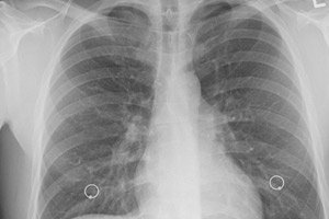
Instrumental diagnostics uses functional pulmonary tests (spirometry and oximetry), X-rays and computed tomography of the chest (CT). In doubtful cases, endoscopic bronchoscopy with lung tissue biopsy is needed. [ 12 ]
Computed tomography provides much more detailed information about changes in the lungs than conventional radiography, and pneumonitis on CT of the lungs is visualized as varying degrees of increase in the thickness of the alveolar walls and the partitions between them. At the same time, the opacity and compaction of the interstitium resembles ground glass, and the pattern of the lungs resembles honeycomb cells (due to small foci of fibrosis).
Differential diagnosis
Hypersensitivity pneumonitis may resemble some infectious and fibrotic lung diseases. Therefore, differential diagnosis of pneumonitis is carried out with obliterating bronchiolitis, bronchial asthma and bronchiectasis; infectious interstitial pneumonia and pneumoconiosis; idiopathic fibrosis, hemosiderosis and alveolar proteinosis of the lungs; granulomatous lung diseases (sarcoidosis, berylliosis, mycobacterial infections), Churg-Strauss syndrome; carcinomatous lymphangitis and sarcoidosis. [ 13 ], [ 14 ]
In many cases, pneumonitis and alveolitis are considered synonyms, for example, allergic alveolitis and hypersensitivity (allergic) pneumonitis are, by all parameters, the same disease. [ 15 ]
Pneumonia or pneumonitis in coronavirus covid?
The cause of COVID-19 is infectious, caused by the SARS-CoV-2 virus. The most common complication is viral interstitial pneumonia with a high probability of developing acute respiratory distress syndrome and subsequent respiratory failure.
At the same time, pneumonia with coronavirus covid has similar symptoms and CT results of the lungs with acute hypersensitivity pneumonitis and immune pneumonitis (associated with cancer treatment with immune checkpoint inhibitors), which complicates diagnosis without thorough testing for the CoV-2 virus.
Pneumonia in COVID-19 is characterized by fever and cough, and respiratory distress syndrome develops later. In pneumonitis, shortness of breath and cough appear immediately, but fever is extremely rare.
More information in the material - Coronavirus infection (atypical pneumonia): causes, symptoms, diagnosis, treatment
Who to contact?
Treatment pneumonitis
Most often, treatment of pneumonitis involves the use of systemic corticosteroids that promote immunosuppression. Oral GCS Prednisolone or Methylprednisolone are prescribed (standard dosage is 0.5 mg/kg of body weight for two to four weeks. Long-term use of corticosteroids increases the risk of infections and can lead to osteoporosis.
The immunosuppressants Mycophenolate mofetil (Supresta, MMF-500), Anakinra (Kineret), Pirfenidone (Esbriet) reduce the formation of antibodies. Side effects of Anakinra include headache, leukopenia and thrombocytopenia. The immune-suppressing drug Pirfenidone is contraindicated in liver and kidney failure. And among its side effects, the instructions indicate headache and dizziness; nausea, vomiting and diarrhea/constipation; loss of appetite and weight; pain in the hypochondrium, joints and muscles; hyperemia of the skin with rashes and itching. [ 16 ]
Other medications are also used, including the fibroblast growth factor receptor and transforming growth factor receptor inhibitor Nintedanib (Vargatef, Ofev) in capsules for oral administration. This drug can cause nausea, vomiting, diarrhea, abdominal pain, decreased appetite, and increased liver transaminase levels.
Treatment of radiation pneumonitis is carried out with corticosteroids, decongestants and drugs that dilate the bronchi.
Breathing problems require oxygen therapy, and in severe cases, artificial ventilation. [ 17 ]
For patients with progressive hypersensitivity pneumonitis, when conservative therapy is ineffective and there is a risk of fatal respiratory failure, surgical treatment is indicated - lung transplantation.
Prevention
Hypersensitivity pneumonitis can be prevented by avoiding known irritants - protecting the airways from dust during work by using a respirator.
But in many cases, if the antigen is not identified, respiratory exposure prevention is problematic.
Forecast
The stage and severity of pneumonitis determine its prognosis. In the mild form of acute hypersensitivity pneumonitis, lung function is most often restored after treatment. And the chronic form of the disease leads to fibrosis, the terminal stage of which can end in severe respiratory failure and, ultimately, death (in almost 60% of cases).