
All iLive content is medically reviewed or fact checked to ensure as much factual accuracy as possible.
We have strict sourcing guidelines and only link to reputable media sites, academic research institutions and, whenever possible, medically peer reviewed studies. Note that the numbers in parentheses ([1], [2], etc.) are clickable links to these studies.
If you feel that any of our content is inaccurate, out-of-date, or otherwise questionable, please select it and press Ctrl + Enter.
Pelvic girdle
Medical expert of the article

Last reviewed: 04.07.2025
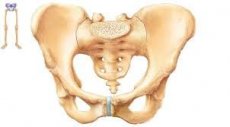
The bones of the pelvic girdle are connected to each other in front by the pubic semi-joint, and at the back with the sacrum they form the sacroiliac joints.
The sacroiliac joint is formed by the auricular surfaces of the sacrum and ilium and is a flat joint. The joint capsule is reinforced by strong short ligaments in front and behind. The sacroiliac interosseous ligament, stretched between the iliac tuberosity and the sacral tuberosity, plays a major role in strengthening the joint. Movements in the joint are insignificant and occur around this ligament, which acts as the axis of the joint. Mobility in the sacroiliac joint provides an elastic buffer function for the pelvis. Branches of the lumbosacral nerve plexus participate in the innervation of the joint. A feature of this joint is the absence of muscles that would specifically set this joint in motion.
The pubic joint (symphysis pubis) is formed by the pubic bones, which are firmly connected to the fibrocartilaginous interpubic disc located between them. Among the pelvic ligaments, it is necessary to note the iliac-lumbar, sacrotuberous and sacrospinous ligaments.
In a vertical position of the body, the pelvis always has an anterior tilt, which is measured by the pelvic tilt angle. It is formed by a line passing through the promontory of the spine and the upper edge of the pubic symphysis, and a line located in the horizontal plane.
This angle is usually 50-60° and can change with changes in the nature of the standing.
During the examination, the physician must remember that the pelvis, together with the presacral disc, pubic and paired or sacral joints, hip joints and muscular-ligamentous apparatus, is a buffer for the movement of the kinematic chain "spine-legs".
There are three main types of pelvis (Lewit K., 1993):
- average type (normal) of the pelvis. The angle of inclination of the sacrum to the vertical from the body's CCP is 130-145°, the vertical passes through the top of the muscles behind the axis of the hip joints. Lumbar lordosis is 18 mm;
- assimilated or liberated pelvis with an elongated sacrum and a high promontory, the lumbosacral disc is higher than L1-L2. The sacrum approaches the vertical line, the angle of inclination is 150-165°, the lumbar lordosis is flattened to 6 mm. Greater mobility of the L1 vertebra and sacroiliac joints is noted;
- The "overloaded" pelvis has a deep-set and forward-protruding promontory. The angle of the sacrum approaches the horizontal line, reaching 110-130°. The plumb line C 7 passes in front of the promontory and the axis of the hip joints. The patient's head is most often pushed forward, the pelvis is pushed back. The structures of the lumbosacral PDS, sacroiliac joint and hip joints are overloaded, the abdominal muscles are overstretched. Hyperlordosis (30 mm) is combined with tonic tension of the multifidus and gluteal muscles. With an overloaded
pelvis, lumbo- and iliosacral joint blockades, interspinous ligamentoses and apiarthrosis (Baastrup syndrome) often occur.
 [
[ Planes and axes of pelvic motion
To study and record the state of the human body and its parts, it is customary to distinguish between body planes and axes of movement. There are three main planes.
The sagittal, or anteroposterior (imaginary) plane divides the human body or any part of it into left and right halves (sections), and the sagittal plane passing through the middle of the body is called the median plane.
The horizontal plane crosses the body transversely, dividing it into the head (cranial) and tail (caudal) sections. The horizontal plane drawn on any limb divides it into the proximal (closer to the body) and distal (further from the body) sections.
The frontal (parallel to the forehead) plane divides the body or its parts into anterior (ventral) and posterior (dorsal) sections. All three planes are perpendicular to each other. Any other plane can only be intermediate in relation to the mentioned planes.
All three planes, when intersecting each other, form lines called axes of rotation. When the sagittal and horizontal planes intersect, the sagittal axis is formed and movement around this axis occurs in the frontal plane. When the frontal and horizontal planes intersect, the transverse axis is formed. Movement around this axis occurs in the sagittal plane. When the sagittal and frontal planes intersect, the vertical axis is formed. Movement around the vertical axis occurs in the horizontal plane.
Biomechanics considers the human movement apparatus as controlled biokinetic chains consisting of links connected to each other by joints and muscles attached to them. Together they form a biomechanism capable of performing specified movements. In a biokinetic chain, movements can be preserved in all joints, only in some of them, or these can be movements of all links as a single whole. Biokinetic chains can be open or closed (with connected end links) and in this regard have different properties. Thus, a closed biokinetic chain does not have a free end link, isolated movements in only one joint are impossible in it.
Использованная литература