
All iLive content is medically reviewed or fact checked to ensure as much factual accuracy as possible.
We have strict sourcing guidelines and only link to reputable media sites, academic research institutions and, whenever possible, medically peer reviewed studies. Note that the numbers in parentheses ([1], [2], etc.) are clickable links to these studies.
If you feel that any of our content is inaccurate, out-of-date, or otherwise questionable, please select it and press Ctrl + Enter.
Optical coherence tomography
Medical expert of the article

Last reviewed: 04.07.2025
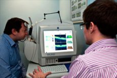
Optical coherence tomography (OCT) is a noninvasive imaging technique that is increasingly used in medicine for diagnostic purposes. An optical coherence tomograph (Humphrey Systems, Dublin, CA) calculates the thickness parameters of the retinal nerve using high-resolution transverse scans of the retina.
 [
[ When is optical coherence tomography used?
Optical coherence tomography is important in detecting glaucoma and monitoring its progression.
Advantages of Optical Coherence Tomography
Optical coherence tomography is of interest for clinical use for a number of reasons. The resolution of OCT is 10–15 μm, which is almost an order of magnitude higher than the resolution of other diagnostic methods, including ultrasound. Such high resolution allows studying tissue architecture. Information obtained using OCT is intravital and reflects not only the structure, but also the features of the functional state of tissues. Optical coherence tomography is noninvasive, since it uses radiation in the near infrared range with a power of about 1 mW, which does not have a damaging effect on the body. The method excludes trauma and does not have the limitations inherent in traditional biopsy.
Who to contact?
How does optical coherence tomography work?
Optical coherence tomography uses a low-coherence interferometer to obtain high-resolution images. The procedure for performing optical coherence tomography is similar to that of ultrasound B-scanning or radar, except that the light is used more as acoustic waves than as radio waves. Distance and microstructure measurements in optical coherence tomography are based on measuring the transit time of light reflected from various microstructural elements of the eye. Sequential longitudinal measurements (A-scanograms) are used to construct a spectrozonal topographic image of tissue microsections that closely resemble histological sections. The resolution of a longitudinal section in optical coherence tomography is about 10 μm, and the resolution of a transverse section is approximately 20 μm. In the clinical evaluation of glaucoma, optical coherence tomography produces cylindrical sections of the retina by scanning a 3.4 mm diameter circle centered at the optic disc. The cylinder is unrolled, presenting a flat cross-sectional image. Optical coherence tomography is used to create a macular thickness map from a series of six radial images traversing the meridians of a clock face, centered at the fovea; the optic disc is similarly mapped, centered at the optic disc, with the radial images. An automated computer algorithm measures the SNL thickness without user intervention. Unlike confocal scanning laser ophthalmoscopy, optical coherence tomography does not require a base plane. The SNL thickness is an absolute cross-sectional parameter. Refraction or axial length of the eye do not affect the optical coherence tomography measurements. The parameters of optical coherence tomography of the thickness of the SNV are independent of tissue birefringence.
How is optical coherence tomography performed?
OCT uses near infrared light to illuminate the tissue area being examined. Any biological tissue, including skin and mucous membranes, consists of structures of varying density and is therefore optically heterogeneous. Infrared light, when it hits the boundary of two media with different densities, is partially reflected from it and scattered. By analyzing the coefficient of backscattering of light, it is possible to obtain information about the structure of the tissue in a given area.
By scanning the tissue with an optical beam, a series of axial measurements are taken in various cross-sections and directions - both axial (in depth) and lateral (sideways). A powerful computer built into the OCT system processes the obtained numerical data and draws a two-dimensional image (a kind of "morphological section"), convenient for visual assessment.
Restrictions
Optical coherence tomography requires a nominal pupil diameter of 5 mm, but in practice most patients can undergo optical coherence tomography without mydriasis. Optical coherence tomography is limited in cortical and posterior subcapsular cataracts.