
All iLive content is medically reviewed or fact checked to ensure as much factual accuracy as possible.
We have strict sourcing guidelines and only link to reputable media sites, academic research institutions and, whenever possible, medically peer reviewed studies. Note that the numbers in parentheses ([1], [2], etc.) are clickable links to these studies.
If you feel that any of our content is inaccurate, out-of-date, or otherwise questionable, please select it and press Ctrl + Enter.
Neck muscles
Medical expert of the article

Last reviewed: 04.07.2025

The muscles and fascia of the neck have a complex structure and topography, which is due to their different origins, different functions, relationships with the internal organs of the neck, blood vessels and nerves. The muscles of the neck are divided into separate groups according to their origin and topographic features (by neck regions).
A distinction is made between muscles that developed on the basis of the first (mandibular) and second (hyoid) visceral gill arches, and muscles that developed from the ventral parts of the myotomes.
The derivatives of the mesenchyme of the first visceral arch are the mylohyoid muscle, the anterior belly of the digastric muscle. The stylohyoid muscle, the posterior belly of the digastric muscle, and the subcutaneous muscle of the neck develop from the mesenchyme of the second visceral arch. The sternocleidomastoid and trapezius muscles are formed from the mesenchyme of the branchial arches. The sternohyoid, sternothyroid, thyrohyoid, omohyoid, geniohyoid, anterior, middle, and posterior scalene muscles, as well as the prevertebral muscles (longus colli and longus capitis) develop from the ventral part of the myotomes. Topographically, the muscles of the neck are divided into superficial and deep.
 [
[ Superficial muscles of the neck
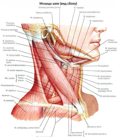
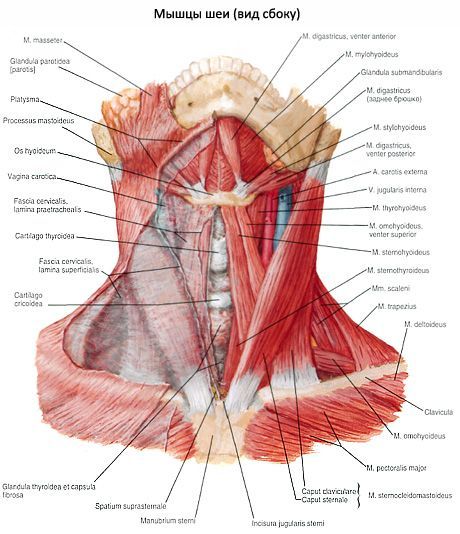
The superficial muscles of the neck include the platysma, sternocleidomastoid, and muscles attached to the hyoid bone, as well as the suprahyoid and infrahyoid muscles. The suprahyoid group of muscles includes the mylohyoid, digastric, stylohyoid, and geniohyoid muscles. The infrahyoid muscles include the sternohyoid, sternothyroid, thyrohyoid, and omohyoid muscles. The deep muscles of the neck are in turn subdivided into the lateral and prevertebral groups. The lateral group includes the anterior, middle, and posterior scalene muscles, which lie to the side of the spinal column. The prevertebral group, located in front of the spinal column, includes the muscles of the head: the anterior rectus capitis, lateral rectus capitis, and the longus colli.
The subcutaneous muscle of the neck (platysma) is thin, flat, and lies directly under the skin. It begins in the thoracic region below the clavicle on the superficial plate of the pectoral fascia, passes upward and medially, occupying almost the entire anterolateral region of the neck. A small area, shaped like a triangle above the jugular notch of the sternum, remains uncovered by the muscle.
Subcutaneous muscle of the neck
The sternocleidomastoid muscle (m. sternocleidomastoideus) is located under the platysma muscle of the neck; when the head is turned to the side, its contour is indicated by a distinct ridge on the anterolateral surface of the neck. This muscle begins with two parts (medial and lateral) on the anterior surface of the manubrium of the sternum and the sternal end of the clavicle. Rising upward and backward, the muscle is attached to the mastoid process of the temporal bone and the lateral segment of the superior nuchal line of the occipital bone. Above the clavicle, between the medial and lateral parts of the muscle, there is a small supraclavicular fossa (fossa supraclavicularis minor).
Muscles attached to the hyoid bone
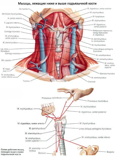
There are muscles located above the hyoid bone - the suprahyoid muscles (mm. suprahyoidei), and muscles located below the hyoid bone - the infrahyoid muscles (mm.infrahyoidei). Both groups of muscles (paired) act on the hyoid bone, which is a support for the muscles involved in important functions: chewing, swallowing, speech, etc. The hyoid bone is held in its position exclusively by the interaction of muscles that approach it from different sides.
Muscles attached to the hyoid bone
Deep muscles of the neck
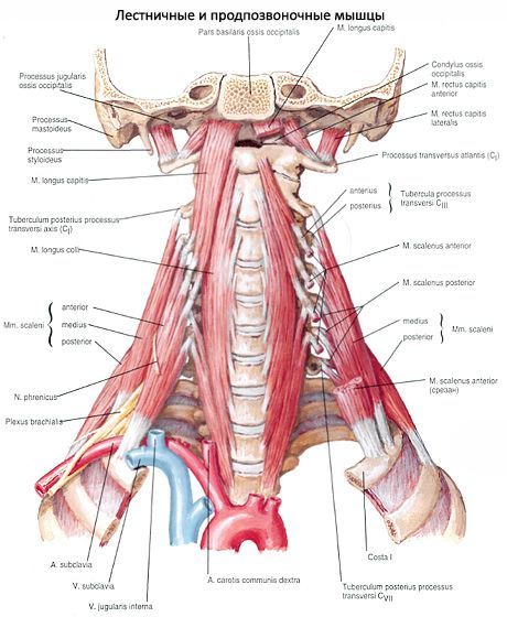
The deep muscles of the neck are divided into lateral and medial (prevertebral) groups.
The lateral group is represented by three scalene muscles. According to their location, the anterior, middle and posterior scalene muscles are distinguished.
Использованная литература