
All iLive content is medically reviewed or fact checked to ensure as much factual accuracy as possible.
We have strict sourcing guidelines and only link to reputable media sites, academic research institutions and, whenever possible, medically peer reviewed studies. Note that the numbers in parentheses ([1], [2], etc.) are clickable links to these studies.
If you feel that any of our content is inaccurate, out-of-date, or otherwise questionable, please select it and press Ctrl + Enter.
The muscles surrounding the opening of the mouth
Medical expert of the article

Last reviewed: 07.07.2025
There are several well-defined muscles around the mouth opening. These muscles include the orbicularis oris, the depressor anguli oris, the depressor labii inferioris, the mentalis and buccinator muscles, the levator labii superioris, the zygomaticus minor and major, the levator anguli oris, and the laughter muscle.
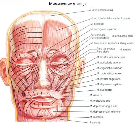
The orbicularis oris muscle forms the muscular basis of the upper and lower lips. This muscle consists of marginal and labial parts, the bundles of which have different orientations.
The marginal part (pars marginalis) is the peripheral, wider section of the muscle. This part is formed by muscle bundles that approach the upper and lower lips from other facial muscles closest to the mouth opening. The marginal part is formed by bundles of the buccinator muscle; the muscle that raises the upper lip; the muscle that raises the corner of the mouth; the muscle that lowers the lower lip; the muscle that lowers the corner of the mouth, etc.
The labial part (pars labialis) is located in the thickness of the upper and lower lips. Bundles of muscle fibers extend from one corner of the mouth to the other.
Both parts (marginal and labial) of the upper and lower lips are woven into the skin and mucous membrane, and also connect with each other in the area of the corners of the mouth and pass from the lower lip to the upper and vice versa.
Function: the orbicularis oris muscle narrows and closes the oral slit and is involved in the act of sucking and chewing.
Innervation: facial nerve (VII).
Blood supply: superior and inferior labial and mental arteries.
The muscle that lowers the angle of the mouth (m.depressor anguli oris) begins at the base of the lower jaw, between the chin and the level of the first premolar. The fibers of this muscle, converging, pass upward and are attached to the skin of the corner of the mouth. At the place of origin of the muscle that lowers the angle of the mouth, part of its bundles intertwine with bundles of the subcutaneous muscle of the neck.
Function: Pulls the corner of the mouth downward and laterally.
Innervation: facial nerve (VII).
Blood supply: inferior labial and mental arteries.
Blood supply: inferior labial and mental arteries.
The muscle that depresses the lower lip (m.depressor labii inferioris) originates at the base of the lower jaw, below the mental foramen. It is partially covered by the muscle that depresses the angle of the mouth. The bundles of the muscle that depresses the lower lip pass upward and medially and are attached to the skin and mucous membrane of the lower lip.
Function: pulls the lower lip downwards and somewhat laterally, acting together with the muscle of the same name on the opposite side, can turn the lip outwards; participates in the formation of expressions of irony, sadness, disgust.
Innervation: facial nerve (VII).
Blood supply: inferior labial and mental arteries.
The mentalis muscle (m.mentalis) is represented by a cone-shaped bundle of muscle fibers that begin on the alveolar eminences of the lateral and medial incisors of the lower jaw, pass downward and medially, connect with the fibers of the same muscle on the opposite side and attach to the skin of the chin.
Function: pulls the skin of the chin upward and laterally (dimples appear on the skin); promotes protrusion of the lower lip forward.
Innervation: facial nerve (VII).
Blood supply: inferior labial and mental arteries.
The buccinator muscle is thin, quadrangular in shape, and forms the muscular base of the cheek. It begins on the oblique line on the branch of the lower jaw and the outer surface of the alveolar arch of the upper jaw at the level of the large molars, as well as on the anterior edge of the pterygomandibular suture, which passes between the lower jaw and the pterygoid hook. The muscle bundles are directed toward the corner of the mouth, partially cross, and continue into the thickness of the muscular base of the upper and lower lips. At the level of the upper molar, the muscle is penetrated by the parotid duct (duct of the parotid salivary gland).
Function: Pulls the corner of the mouth back; presses the cheek against the teeth.
Innervation: facial nerve (VII).
Blood supply: buccal artery.
The muscle that raises the upper lip (m. levator labii superioris) begins on the entire infraorbital edge of the upper jaw. The muscle bundles converge downwards and are woven into the thickness of the corner of the mouth and into the wing of the nose.
Function: raises the upper lip; participates in the formation of the nasolabial fold, which extends from the lateral side of the nose to the upper lip; pulls the wing of the nose upward.
Innervation: facial nerve (VII).
Blood supply: infraorbital and superior labial arteries.
The zygomaticus minor muscle (m.zygomaticus minor) originates on the zygomatic bone at the lateral edge of the muscle that raises the upper lip. Bundles of the zygomaticus minor muscle pass downward medially and intertwine in the skin of the corner of the mouth.
Function: Raises the corner of the mouth.
Innervation: facial nerve (VII).
Blood supply: infraorbital and buccal arteries.
The zygomaticus major muscle (m.zygomaticus major) originates on the zygomatic bone and is attached to the corner of the mouth.
Function: pulls the corner of the mouth outward and upward, is the main muscle of laughter.
Innervation: facial nerve (VII).
Blood supply: infraorbital and buccal arteries.
The muscle that raises the angle of the mouth (m.levator anguli oris) originates on the anterior surface of the upper jaw in the area of the canine fossa; it is attached to the corner of the mouth.
Function: pulls the angle of the upper lip upward and laterally.
Innervation: facial nerve (VII).
Blood supply: infraorbital artery.
The laughter muscle (m.risorius) originates on the chewing fascia, runs forward and medially, and is attached to the skin of the corner of the mouth. It is usually poorly expressed and often absent.
Function: pulls the corner of the mouth laterally, forming a dimple on the cheek.
Innervation: facial nerve (VII).
Blood supply: facial artery, transverse cervical artery.
 [
[