
All iLive content is medically reviewed or fact checked to ensure as much factual accuracy as possible.
We have strict sourcing guidelines and only link to reputable media sites, academic research institutions and, whenever possible, medically peer reviewed studies. Note that the numbers in parentheses ([1], [2], etc.) are clickable links to these studies.
If you feel that any of our content is inaccurate, out-of-date, or otherwise questionable, please select it and press Ctrl + Enter.
Shoulder girdle muscles
Medical expert of the article

Last reviewed: 07.07.2025
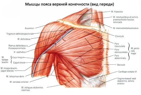
The deltoid muscle (m.deltoideus) is located superficially, directly under the skin, covers the shoulder joint from the lateral side, from the front, from above and from behind, forms the characteristic roundness of the shoulder). This muscle is separated from the pectoralis major by the deltoid-pectoral groove (sulcus deltoideopectoralis). The deltoid muscle has a pennate structure and an extensive origin. It begins on the anterior edge of the lateral third of the clavicle, the outer edge of the acromion, on the spine of the scapula and the adjacent part of the infraspinatus fascia. Accordingly, three parts of the deltoid muscle are distinguished: clavicular, acromial and scapular. The bundles of all three parts of the muscle converge on the outer surface of the humerus and are attached to the deltoid tuberosity.
The supraspinatus muscle (m.supraspinatus) is located in the supraspinous fossa. It originates on the posterior surface of the scapula above the scapular spine and on the supraspinatus fascia. The bundles extend laterally. The muscle is attached to the upper surface of the greater tubercle of the humerus; some of the bundles of the supraspinatus muscle are woven into the capsule of the shoulder joint. The infraspinatus muscle (m.infraspinatus) originates on the posterior surface of the scapula below its spine and on the fascia of the same name. The bundles of the muscle converge and extend laterally and slightly upward (behind the shoulder joint). The muscle is attached to the middle surface of the greater tubercle of the humerus. Some of the bundles are woven into the capsule of the shoulder joint.
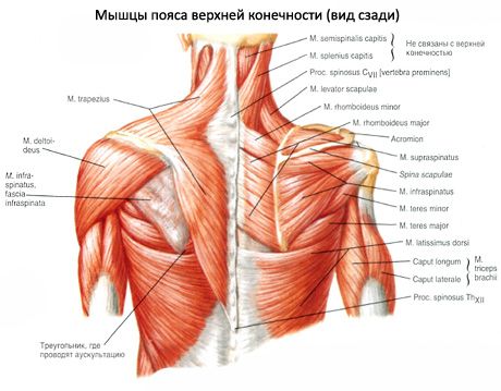
Supraspinatus and infraspinatus muscles
The small teres muscle (m.terpes minor) originates on the lateral edge of the scapula and the infraspinatus fascia; it is attached to the lower surface of the greater tubercle of the humerus. It is directly adjacent below to the infraspinatus muscle, and is covered from behind by the scapular part of the deltoid muscle. The large teres muscle (m.terpes major) originates on the lower part of the lateral edge and lower angle of the scapula, on the infraspinatus fascia.
The muscle bundles are directed medially and upward along the lateral edge of the scapula, crossing the humerus on the medial side below the level of its surgical neck. They are attached by a wide flat tendon to the crest of the lesser tubercle of the humerus, distal and somewhat posterior to the attachment of the tendon of the latissimus dorsi.
Teres minor and teres major muscles
The subscapularis muscle (m. subscapularis) is wide, thick, triangular in shape. It occupies almost the entire costal surface of the scapula. It has a fleshy origin on the surface of the subscapular fossa and the lateral edge of the scapula. It is attached by a flat tendon to the lesser tubercle and the crest of the lesser tubercle of the humerus. At the point of attachment between the tendon and the capsule of the shoulder joint, there is a subtendinous bursa of the subscapularis muscle, which usually communicates with the cavity of the shoulder joint.
 [
[ How to examine?