
All iLive content is medically reviewed or fact checked to ensure as much factual accuracy as possible.
We have strict sourcing guidelines and only link to reputable media sites, academic research institutions and, whenever possible, medically peer reviewed studies. Note that the numbers in parentheses ([1], [2], etc.) are clickable links to these studies.
If you feel that any of our content is inaccurate, out-of-date, or otherwise questionable, please select it and press Ctrl + Enter.
Pelvic muscles (pelvic girdle muscles)
Medical expert of the article

Last reviewed: 04.07.2025

The pelvic muscles are divided into two groups - internal and external. The internal group of muscles includes the iliopsoas, internal obturator and piriformis. The external group of pelvic muscles includes the gluteus maximus, gluteus medius and gluteus minimus: the tensor of the broad fascia, the quadratus femoris and the external obturator.
 [ 1 ]
[ 1 ]
Internal pelvic muscle group
The iliopsoas muscle (m.iliopsoas) consists of two muscles - the lumbar major and the iliac, which, starting in different places (on the lumbar vertebrae and the ilium), join into a single muscle attached to the lesser trochanter of the femur. Both parts of the muscle participate in the formation of the posterior wall of the abdominal cavity.
The small lumbar muscle (m.psoas minor) is inconstant, absent in 40% of cases. It originates on the intervertebral disc and the adjacent edges of the bodies of the 12th thoracic and 1st lumbar vertebrae. The muscle is located on the anterior surface of the large lumbar muscle, fused with the fascia covering it. The thin belly of this muscle passes into a long tendon, which is attached to the arcuate line of the ilium and to the iliopectineal eminence. Some of the bundles of the tendon of this muscle are woven into the iliac fascia and into the iliopectineal arch.
The internal obturator muscle (m.obturatorius internus) originates at the edges of the obturator foramen (except for the obturator groove), on the internal surface of the obturator membrane, on the pelvic surface of the ilium (above the obturator foramen) and on the obturator fascia. The internal obturator muscle exits the pelvic cavity through the lesser sciatic foramen, changes direction at an acute angle, and goes over the edge of the lesser sciatic notch (there is a sciatic bursa of the internal obturator muscle, bursa ischiadica m.obturatorii interni).
The piriformis muscle (m piriformis) originates on the pelvic surface of the sacrum (II-IV sacral vertebra), lateral to the pelvic sacral openings, and exits the cavity of the lesser pelvis through the greater sciatic opening. Behind the neck of the femur, the muscle passes into a round tendon that attaches to the apex of the greater trochanter. Under this muscle is the synovial bursa of the piriformis muscle (bursa synovialis musculi piriformis).
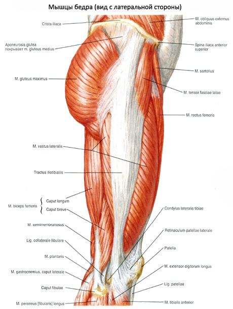
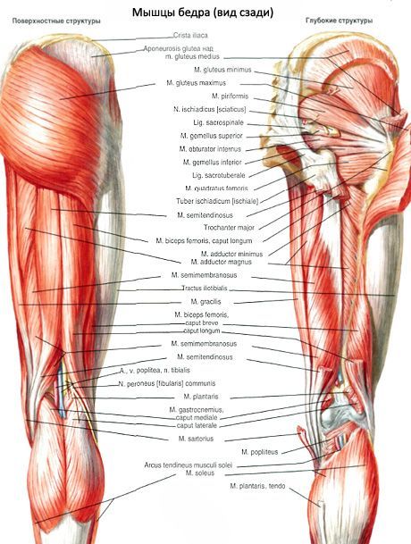
External pelvic muscle group
The external pelvic muscles are located in the gluteal region and on the lateral surface of the pelvis. Having relatively large surfaces of the origin of the muscles on the bones of the pelvic girdle, the bundles of these muscles follow in the direction of their attachment on the femur. The external pelvic muscles form 3 layers: superficial, middle and deep.
The superficial layer consists of the gluteus maximus and the tensor fasciae latae. The middle layer contains the gluteus medius and the quadratus femoris. This group includes the extrapelvic parts of the piriformis and internal obturator muscles, the superior and inferior gemellus muscles. The deep layer includes the gluteus minimus and external obturator muscles. All of these muscles act on the hip joint.
The gluteus maximus (m.gluteus maximus) is strong, has a large-bundle structure, and stands out in relief due to its large mass in the gluteal region (regio glutea). This muscle reaches its greatest development in humans due to upright posture. Situated superficially, it has a wide origin on the ilium (linea glutea posterior), on the initial (tendon) part of the muscle that straightens the spine, on the dorsal surface of the sacrum and coccyx, on the sacrotuberous ligament.
The gluteus medius muscle (m.gluteus medius) originates on the gluteal surface of the ilium, between the anterior and posterior gluteal lines, on the broad fascia. The muscle goes down, passes into a thick tendon, which is attached to the top and outer surface of the greater trochanter.
The gluteus minimus (m.gluteus minimus) is located under the gluteus medius. It originates on the outer surface of the iliac wing between the anterior and inferior gluteal lines, along the edge of the greater sciatic notch. It is attached to the anterolateral surface of the greater trochanter of the femur; some of the bundles are woven into the capsule of the hip joint. Between the tendon of the muscle and the greater trochanter there is a trochanteric bursa of the gluteus minimus (bursa trochanterica musculi glutei minimi).
The tensor fasciae latae originates on the anterior superior iliac spine and the adjacent part of the iliac crest. The muscle is located between the superficial and deep plates of the fascia lata. At the level of the border between the upper and middle thirds of the thigh, the muscle passes into the iliotibial tract of the fascia lata (tractus iliotibialis), which continues downward and attaches to the lateral condyle of the tibia.
The quadratus femoris is a flat, quadrangular muscle located between the inferior gemellus muscle at the top and the upper edge of the adductor magnus at the bottom. It begins at the upper part of the outer edge of the ischial tuberosity and is attached to the upper part of the intertrochanteric crest. There is often a synovial bursa between the anterior surface of the muscle and the greater trochanter.
The external obturator muscle (m.obturatorius externus) is triangular in shape, originates on the outer surface of the pubic bone and the branch of the ischium, as well as on the medial two-thirds of the obturator membrane. The muscle bundles converge and are directed backward, laterally and upward. The tendon of the muscle passes behind the hip joint and is attached to the trochanteric fossa of the femur and to the joint capsule.
Использованная литература