
All iLive content is medically reviewed or fact checked to ensure as much factual accuracy as possible.
We have strict sourcing guidelines and only link to reputable media sites, academic research institutions and, whenever possible, medically peer reviewed studies. Note that the numbers in parentheses ([1], [2], etc.) are clickable links to these studies.
If you feel that any of our content is inaccurate, out-of-date, or otherwise questionable, please select it and press Ctrl + Enter.
Muscles of the foot
Medical expert of the article

Last reviewed: 04.07.2025

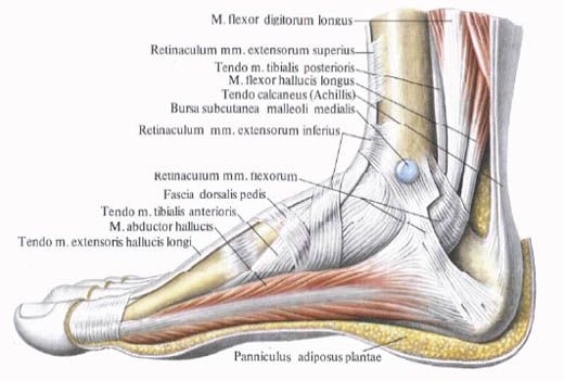
Along with the tendons of the lower leg muscles attached to the bones of the foot, which are part of the anterior, posterior and lateral groups, the foot has its own (short) muscles.
These muscles originate and are attached within the skeleton of the foot, have complex anatomical-topographical and functional relationships with the tendons of those muscles of the lower leg, the attachment points of which are located on the bones of the foot. The muscles of the foot are located on its dorsum and on the sole.
 [
[ Muscles of the dorsum of the foot
The muscles of the dorsum of the foot lie beneath the dorsal fascia of the foot and the tendons of the long extensors of the fingers. These two muscles are the short extensor of the fingers and the short extensor of the big toe.
The short extensor of the fingers (m.extensor digitorum brevis) is a poorly developed muscle. It originates on the anterosuperior and lateral surfaces of the calcaneus. The muscle passes along the dorsal surface of the foot obliquely forward and medially. Three tendons of this muscle reach the II-IV toes, join on the lateral side to the tendons of the long extensor of the fingers and together with them are attached to the bases of the middle and distal phalanges.
Function: Together with the tendons of the long extensor of the fingers, it participates in the extension of the toes.
Innervation: deep peroneal nerve (LIV-SI).
Blood supply: lateral tarsal and peroneal arteries.
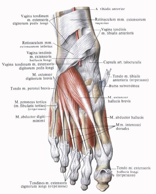
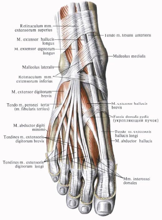
The short extensor of the big toe (m.extensor hallucis brevis) lies medial to the short extensor of the fingers. It begins on the upper surface of the calcaneus, in its anterior part. The muscle is directed forward and medially, passes into a tendon that is attached to the dorsal surface of the base of the proximal phalanx of the big toe.
Function: Participates in the extension of the big toe.
Innervation: deep peroneal nerve (LIV-SI).
Blood supply: dorsalis pedis artery.
Muscles of the sole of the foot
In the area of the sole of the foot, the following muscle groups are distinguished: medial - on the side of the big toe, lateral - on the side of the little toe, middle, occupying an intermediate position.
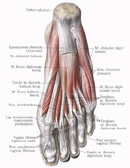
Unlike the hand, the medial and lateral groups on the sole of the foot are represented by a smaller number of muscles, and the middle group is strengthened. In total, there are 14 short muscles on the sole. Three of them belong to the medial group (the muscle that abducts the big toe, the short flexor of the big toe, and the muscle that adducts the big toe). Two muscles form the lateral group (the muscle that abducts the little toe and the short flexor of the little toe). The middle group on the sole is strengthened. It includes 13 muscles. In addition to 4 lumbrical and 7 interosseous muscles, it includes two more muscles - the short flexor of the fingers and the square muscle of the plant.
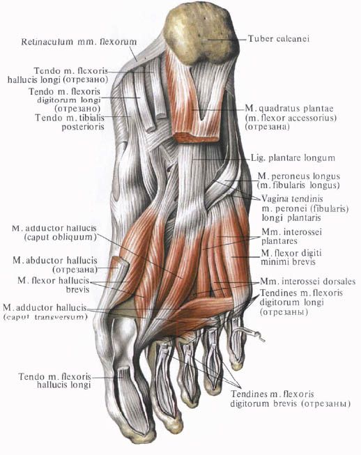
Medial group of muscles of the sole of the foot
The muscle that abducts the big toe (m.abductor hallucis) lies superficially along the medial edge of the foot. It begins with short tendinous bundles on the medial surface of the calcaneal tuberosity, fleshy bundles - on the inferior retinaculum of the flexor tendons and the plantar aponeurosis. The muscle is attached to the medial side of the base of the proximal phalanx of the big toe.
Function: Abducts the big toe from the midline of the sole of the foot in a medial direction.
Innervation: medial plantar nerve (LV-SI).
Blood supply: medial plantar artery.
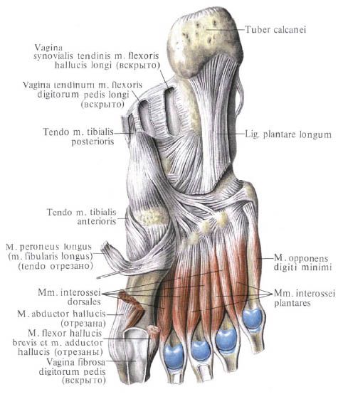
The short flexor of the big toe (m.flexor hallucis brevis) adjoins the previous muscle on the lateral side. It begins with a narrow tendinous plate on the medial side of the plantar surface of the cuboid bone (behind the groove of the tendon of the long peroneus muscle), on the first cuneiform bone and the plantar calcaneocuboid ligament. The muscle goes forward and divides into medial and lateral parts, between which the tendon of the long flexor of the big toe passes.
Both parts of the muscle are attached to the base of the proximal phalanx and to the sesamoid bones on either side of the first metatarsophalangeal joint. On the lateral side, the muscle is fused with the adductor hallucis muscle.
Function: flexes the big toe.
Innervation: lateral part of the muscle - lateral plantar nerve (SI-SII); medial part - medial plantar nerve (LV-SI).
Blood supply: medial plantar artery, plantar arch.
The adductor hallucis muscle lies deep, almost in the middle of the sole. It has two heads: oblique and transverse. The oblique head (caput obliquum) originates on the cuboid, lateral cuneiform, at the base of the II, III and IV metatarsal bones and on the long plantar ligament. The muscle belly is directed forward and medially, passing into a common tendon with the transverse head. The transverse head (caput transversum) forms a narrow flat muscle belly, which originates on the capsules of the metatarsophalangeal joints of the III-V toes, goes transversely in the medial direction and connects with the oblique head. The tendon of the adductor muscle is attached to the base of the proximal phalanx of the big toe and to the lateral sesamoid bone.
Function: brings the big toe to the midline of the foot, participates in flexion of the big toe.
Innervation: lateral plantar nerve (SI-SII).
Blood supply: plantar metatarsal arteries, plantar arch.
Lateral group of muscles of the sole of the foot
The muscle that abducts the little toe of the foot (m.abductor digiti minimi) begins with tendon and muscle bundles on the plantar surface of the calcaneus, the tuberosity of the 5th metatarsal bone and on the plantar aponeurosis. The tendon of the muscle runs along the lateral edge of the foot and is attached to the lateral side of the proximal phalanx of the little toe.
Function: flexes the proximal phalanx of the little finger and abducts it laterally.
Innervation: lateral plantar nerve (SI-SII.
Blood supply: lateral plantar artery.
The short flexor of the little toe of the foot (m.flexor digiti minimi brevis) originates on the medial side of the plantar surface of the V metatarsal bone, on the sheath of the tendon of the long peroneus muscle and on the long plantar ligament. The tendon of the muscle lying more medial and deeper than the previous one is attached to the base of the proximal phalanx of the little toe.
Function: bends the little finger.
Innervation: lateral plantar nerve (SI-SII.
Blood supply: lateral plantar artery.
The opposing muscle of the little finger (m.opponens digiti minimi) is located on the lateral side of the short flexor of the little finger. It begins on the long plantar ligament. It is attached to the V metatarsal bone.
Function: Participates in strengthening the lateral longitudinal arch of the foot. The muscle is not permanent.
Innervation: lateral plantar nerve (SI-SII).
Blood supply: lateral plantar artery.
Middle group of muscles of the sole of the foot
The short flexor of the fingers (m.flexor digiti brevis) lies under the plantar aponeurosis. On the lateral side, the muscle is adjacent to the muscle that abducts the little finger, and on the medial side - to the muscle that abducts the big toe. Under the short flexor of the fingers are the quadratus plantaris muscle and the tendons of the long flexor of the fingers. The short flexor of the fingers originates on the anterior part of the plantar surface of the calcaneal tubercle and on the plantar aponeurosis. From the flat muscle belly of this muscle, 4 tendons extend, which are attached to the middle phalanges of the II-V fingers. Each of these tendons at the level of the proximal phalanx splits into two bundles. The tendon of the long flexor of the fingers passes through the gap between them. Some of the bundles of the tendons of the short flexor of the fingers are woven directly into the fibrous sheaths of the toes. The indicated relationships of the tendons of the short flexor of the fingers with the tendons of the long flexor of the fingers on the foot are similar to the relationships of the tendons of the superficial and deep flexors of the fingers of the hand.
Function: flexes the II-V toes; participates in strengthening the longitudinal arch of the foot.
Innervation: medial plantar nerve (LV-SI).
Blood supply: medial and lateral plantar arteries.
The quadratus plantaris, accessory flexor (m.quadratus plantae, smflexor accessorius) originates on the lateral and medial sides of the lower surface of the calcaneus and on the long plantar ligament. The muscle is directed forward and at the level of the middle of the sole of the foot is attached on the lateral side to the tendons of the long flexor of the fingers, directed to the II-IV toes.
Function: participates in the flexion of the toes, while simultaneously giving the pull of the long flexor of the fingers a straight direction.
Innervation: lateral plantar nerve (SI-SII.
Blood supply: lateral plantar artery.
Lumbrical muscles (mm.lumbricales); there are 4 of them, and they have a fusiform shape. The 3 muscles lying laterally originate on the surfaces of the tendons of the long flexor of the fingers facing each other. The fourth, medially located muscle, originates on the medial side of the adjacent tendon of the long flexor of the fingers. Each lumbrical muscle continues into a thin tendon, which is attached on the medial side to the proximal phalanx of the corresponding finger (II-V). Some of the bundles of tendons of the lumbrical muscles bend around the proximal phalanx and pass to the back of the fingers, intertwining with the tendons of the long extensor of the toes.
Function: flexes the proximal and extends the middle and distal phalanges of the II-V toes, moving them medially towards the big toe.
Innervation: lateral and medial plantar nerves (LV-SI).
Blood supply: lateral and medial plantar arteries.
The interosseous muscles (m.Interossei) are located in the spaces between the metatarsal bones. These muscles are divided into two groups: plantar interosseous and dorsal interosseous muscles.
Unlike the similar muscles located on the hand, which are grouped on the sides of the middle finger, the interosseous muscles on the foot are concentrated on the sides of the second finger. This is due to the specificity of the function: grasping - of the hand and musculoskeletal - of the foot.
Plantar interosseous muscles (mm.interossei plantares); there are 3 of them, located in the interosseous spaces on the side of the sole. Each muscle originates at the base of the medial surface of the bodies of the III-V metatarsal bones. The plantar muscles are attached to the medial surface of the proximal phalanges of the III-V toes. Some of the bundles pass from the medial side to the dorsal surface of the corresponding toe and are intertwined into the dorsal aponeurosis.
Function: The plantar interosseous muscles adduct the III-V toes to the II toe; flex the proximal phalanges of these toes.
Innervation: lateral plantar nerve (SI-SII).
Blood supply: plantar metatarsal arteries, plantar arch.
Dorsal interosseous muscles (mm.interossei dorsales); there are 4 of them, occupying the spaces between the metatarsal bones on the dorsal side. Each dorsal interosseous muscle begins with two heads on the surfaces of adjacent metatarsal bones facing each other. The tendons of the muscles are attached to the base of the proximal phalanges and to the tendons of the long extensor of the fingers. The first interosseous muscle is attached to the medial side of the second toe, the other 3 - to the lateral side of the second-fourth toes.
Function: the first dorsal interosseous muscle abducts the II toe from the midline of the foot toward the big toe. The other 3 muscles (second to fourth) abduct the II-IV toes laterally (bring them closer to the little toe). The dorsal interosseous muscles flex the proximal phalanges of the II-IV toes.
Innervation: lateral plantar nerve (SI-SII).
Blood supply: plantar metatarsal arteries, plantar arch.
Movements of the toes (unlike the fingers) are possible within small limits, mainly around the frontal axis (flexion - extension). The big toe has somewhat greater mobility compared to the other toes.
Flex the big toe: long and short extensors of the big toe.
Adductor hallucis: muscle that adducts the big toe.
Abductor hallucis: muscle that abducts the big toe.
The flexors of the toes longus and shortus flex the second through fifth toes. The extensors of the toes longus and shortus extend these toes.
Использованная литература