
All iLive content is medically reviewed or fact checked to ensure as much factual accuracy as possible.
We have strict sourcing guidelines and only link to reputable media sites, academic research institutions and, whenever possible, medically peer reviewed studies. Note that the numbers in parentheses ([1], [2], etc.) are clickable links to these studies.
If you feel that any of our content is inaccurate, out-of-date, or otherwise questionable, please select it and press Ctrl + Enter.
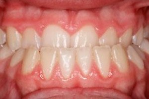
Mesial bite
Medical expert of the article

Last reviewed: 04.07.2025
One of the most uncomfortable deviations of dental development is considered to be mesial bite, which in dentistry is also called progenia, or anterior bite. The pathology is characterized by a clear protrusion of the lower jaw forward. The difficulty lies in the fact that, in addition to the aesthetic problem, such occlusion contributes to the emergence of many health problems. In particular, patients with mesial bite often develop diseases of the digestive tract and oral cavity, sleep disorders, headaches, etc. An unpleasant appearance and incorrect facial geometry can cause many psycho-emotional problems. In this article, we will talk about the features of mesial bite. [ 1 ]
Epidemiology
At the stage of formed bite (this occurs from the age of 17 onwards), problems with the dental mechanism are registered in approximately 35% of people (we mean patients who have not previously been treated for such anomalies). Among all known dental defects, mesial bite occurs in approximately 2-6%. [ 2 ] Among them:
- almost 14% against the background of normal jaw development;
- 19% due to maxillary underdevelopment;
- 25% with excessive growth of the mandibular body and branch;
- 16% with excessive growth of the mandibular body;
- 3% with excessive growth of only the mandibular branch;
- 18% against the background of a combination of all the listed signs.
In older patients, mesial bite of an indefinite form can be diagnosed based on the existing dental and jaw symptoms. Clarification of the form is more difficult and requires additional diagnostic measures.
Causes mesial bite
True mesial bite in almost every second case is a congenital disorder (hereditary defect). The problem may be a consequence of a difficult period of bearing the future baby, or complicated childbirth associated with the child's progression through the birth canal. The true type of bite anomaly can be diagnosed already in the first year of the baby's life. [ 3 ]
However, heredity is not the only root cause of mesial bite: the disease can develop after birth. There are a number of prerequisites for this:
- diseases affecting the upper dentition or upper jaw;
- premature or delayed change of temporary teeth (meaning not only physiological change, but also that associated with traumatic loss of primary teeth);
- bad childhood habits (keeping fingers in the mouth for a long time, using pacifiers and nipples, etc.);
- incorrect position of the child during sleep or at the table (for example, resting the chin on the hand, etc.);
- cranial injuries;
- shortened frenulum of the tongue;
- disorders associated with the skeletal system, rickets;
- otolaryngological diseases, curvature of the nasal bones, etc.
In some patients, the cause may be jaw osteomyelitis, tumor processes, acromegaly, complications after removal of the cleft palate.
Despite the abundance of reasons, it must be admitted that mesial bite after braces can be completely corrected. However, long-term painstaking treatment will be required - usually at least 18 months, and sometimes even more. Therefore, the patient is advised to be patient and strictly follow the advice and instructions of his attending physician.
Risk factors
The appearance of mesial bite is caused by a whole combination of factors that affect the dental mechanism at different stages of its formation. One of the primary causes that determine the development of pathology is heredity. Thus, genetic disorders occur in approximately 40-60% of patients with malocclusion. [ 4 ]
The second category of persistent unfavorable factors affects the intrauterine development of the baby and causes the appearance of specific defects - for example, bone curvatures, underdevelopment of muscles, etc. Disorders of maxillofacial functionality and bad habits also play a role - all these factors significantly increase the risk of developing orthodontic problems.
How can posture affect the quality of the bite? Normal correct position of the body and spine is accompanied by an optimal ratio of the lower and upper jaws, since there is an interaction of the weight vectors of the lower jaw, neck muscles, trachea, back, and oral floor. With adequate distribution of gravity, muscle traction and pressure, the lower jaw is in a position that corresponds to a quality bite, and the bone dental system experiences a load that is feasible for it. If the posture is incorrect, then the resultant action of these forces changes: a mandibular displacement is noted, a mesial bite is formed. An adverse effect is often caused by a night's rest with a soft mattress and a high pillow, placing hands under the head, etc.
Another important factor is impaired nasal breathing. In such a situation, the patient constantly opens his mouth, the oral diaphragm weakens, which leads to aggravation of the lower facial segment, the appearance of a double chin, and a change in the jaw relationship.
In general, doctors talk about the following most common adverse factors:
- heredity (there are relatives in the family with mesial bite or other similar disorders);
- underdevelopment, defects of the dental mechanism;
- bad habits, sucking a pacifier, finger, pencil, upper lip, etc.;
- poor posture or curvature of the spine;
- impaired function of ENT organs, etc.
We will talk in more detail about the negative influences of external and internal factors below.
Pathogenesis
In mesial occlusion, the anterior teeth close in a reverse relationship along the sagittal plane. The depth of such reverse overlap may vary. In particularly complex cases, the cutting edges of the upper anterior teeth contact the mucous tissue of the mandibular alveolar process on the tongue side.
It happens that a patient is diagnosed with both open and mesial bite. The severity of the defect is determined by the size of the sagittal gap. The lateral teeth close in accordance with the third class according to Angle. In the case of a complex course of the pathology, the closure of the first upper and second lower molars is observed. In some cases, the presence of a crossbite (unilateral or bilateral lingual) is noted. [ 5 ]
External symptoms of the defect may have different severity, which depends on the form and degree of complexity. A concave facial profile, a massive protruding chin, a “hidden” upper lip, a high face and an expanded mandibular angle indicate that mesial bite is related to excessive development of the lower jaw.
Taking into account the scale of the discrepancy between the dental arches, specialists have identified several degrees of mesial bite:
- The first degree involves a reverse overlap of the anterior teeth, in which there is mutual contact, or a sagittal gap of up to 2 mm, an increase in the angles of the lower jaw to 1310, an incorrect relationship of the first molars along the sagittal plane of up to 5 mm, and a disrupted localization of individual crowns.
- In the second degree, the width of the sagittal gap is up to 10 mm, the sagittal ratio of the first molars is disturbed up to 10 mm, the angles of the lower jaw are increased up to 1330, the localization of individual crowns is disturbed, and the maxillary narrowing is observed. The simultaneous presence of an open bite is possible.
- In the third degree, the width of the sagittal gap exceeds 1 cm, there are discrepancies in the sagittal ratio of the first molars within 11-18 mm, the mandibular angle is rotated to 145 degrees.
In general, experts talk about the following root causes of mesial bite:
- individual characteristics of the musculoskeletal system that are transmitted by autosomal dominant inheritance (occur in approximately 30% of cases);
- diseases of a woman during pregnancy;
- birth injuries;
- artificial feeding with inferior mixtures;
- diseases of the musculoskeletal system (in particular, rickets);
- bad habits since childhood;
- enlarged tongue, abnormal tongue function, shortened frenulum;
- dental defects;
- enlarged tonsils;
- incorrect sleeping position (chin tucked into chest, etc.);
- incorrect jaw or tooth size;
- maxillary adentia;
- "extra" teeth in the lower row.
Symptoms mesial bite
The clinical picture of mesial occlusion is varied. The first signs – both facial and intraoral – are always less pronounced during the period of temporary teeth than during permanent occlusion.
In case of true mesial bite, the symptoms are represented by a separate symptom complex, which reflects excessive development and specific configuration of the lower jaw.
The upper jaw may be of normal size, short, or distal to the skull: this can be determined by teleradiography. In some patients, the disproportionate position of the jaws is compensated by their mutual arrangement.
Examination of the facial profile reveals an elongation of the mandibular body and an increase in the angle between the ramus and the body. A "sinking" of the middle third of the face is noted, with a protruding chin and lower lip. If the mesial bite is combined with an open bite, the face takes on an elongated appearance, since the dimensions of its lower third increase.
Visual examination reveals inadequate width of the jaw dental arches in the molar and premolar area, a shortened anterior segment of the upper arch, a narrowed and shortened upper apical base, and in some cases, upper incisor retrusion and upper canine retention due to their infringement in the upper arch.
In the anterior part, different types of reverse overlap can be observed - both pronounced open overlap with a sagittal interdental gap and deep overlap.
In general, external symptoms are most often represented by the following signs:
- "concave" face;
- unpleasant sensations and sounds in the temporomandibular joint during chewing, talking, swallowing, etc.;
- protrusion of the lower incisors forward during the apposition of the teeth;
- joint and muscle facial pain;
- widening and retraction of the upper lip;
- speech disorders (lisping, slurring);
- discomfort when biting off pieces of food.
In the absence of qualified medical care, mesial bite in adults causes not only changes in the facial skeleton, but also difficulties with the restoration of crowns (problem treatment, prosthetics). Dental disorders are often associated with increased load, which is placed on the lower dentition. Accelerated abrasion of tooth enamel is observed, gum injuries, gingivitis and other diseases of the oral cavity often occur. To avoid this, mesial bite correction should be done in childhood.
Unfortunately, the vast majority of patients suffering from mesial bite, with age, get used to the discomfort associated with changes in the dentoalveolar apparatus, and practically do not notice the inconvenience. But it is still better to think about consulting a specialist in time and correct the problem in advance. [ 6 ]
Mesial bite in a child
Mesial bite can form in the fetus while it is still in the mother’s womb – this happens as a result of the genetic characteristics of one of the parents (less often – both parents at once).
After the baby is born, the bite can be spoiled by many factors - for example, sucking the upper lip, sleeping with the head lowered to the chest, etc. [ 7 ]
In childhood, unlike in adulthood, the skeletal system is not yet fully formed. In this regard, any impact on the dental mechanism is easier, and the bite is corrected faster and better. If a small correction of the position of the dentition or individual crowns is required, then from about the age of seven, removable vestibular plates are used for treatment. With a more serious degree of mesial bite, the installation of a bracket system may be required. [ 8 ], [ 9 ]
Forms
Mesial bite can be:
- jaw, or skeletal - that is, associated with abnormal bone development;
- dental, or dentoalveolar – caused by the incorrect positioning of the crowns in the alveolar processes.
Depending on the location, mesial bite can be:
- general (misalignment is observed both in the area of the frontal and in the area of the lateral teeth);
- partial (pathology is observed only in the frontal zone).
In addition, there is a bite without mandibular displacement, or with displacement.
According to the etiological features, we speak of true and false progenia. The basis of true mesial bite is the increased size of the mandibular branch and/or body. The false variant is a frontal progenia disorder or forced mesial bite, which develops in the absence of abrasion of the tubercles of the milk mandibular canines against the background of normal jaw rows. In a calm state, the patient does not show pathological signs - until he closes his teeth: the jaw moves forward, reaching the mesial ratio. [ 10 ]
Other possible forms of pathology:
- An open mesial bite, in addition to the forward protrusion of the lower jaw, is characterized by the absence of contact between most of the antagonist crowns (molars or incisors).
- Cross mesial bite is characterized by insufficient development of one side of the dental row. As a result, on one jaw side the lower teeth overlap the upper ones, and on the other side – vice versa.
- The gnathic form of mesial bite is determined by the change in the mandibular angles – up to 145-150.
Complications and consequences
Mesial bite is a pathology of the dentoalveolar mechanism that is prone to relapse. If timely measures to eliminate the defect are not taken, such a pathology can progress, contributing to the development of more complex anomalies and diseases.
One of the most common consequences of mesial bite is disturbed facial proportions and lack of harmonious appearance. The patient has an unpleasant "sunken" profile caused by the protrusion of the lower jaw forward (the so-called "mesial protrusion"). This type of bite can be combined with individual dental or dentoalveolar defects - for example, anterior mandibular displacement can lead to reverse overlap in the area of the frontal crowns.
The presence of a sagittal gap can impair chewing function, since the chewing effect is reduced due to the lingual contact of the anterior teeth.
Impaired chewing, in turn, negatively affects the functioning of the digestive organs, as well as the functional capacity of the temporomandibular joint. Various joint pathologies appear - for example, of an inflammatory or dystrophic nature. [ 11 ]
Severe reverse overlap can lead to chronic damage to the periodontium, which is associated with constant contact of the frontal row of teeth with the mandibular gum. As a result, gingivitis, periodontosis, and periodontitis develop.
A slight reverse overlap (the front teeth are located end to end) often leads to increased abrasion of the crowns. The increased load on the chewing molars is compensated for some time, but after a while destructive processes begin.
Skeletal defect of the third class according to Angle leads to difficulties in carrying out orthopedic and orthodontic treatment manipulations. Patients may have speech and pronunciation disorders. Complaints of temporomandibular pain, radiating to the area of the auricles and head, as well as joint crunching are common. The severity of negative consequences depends on the neglect of such a pathology as mesial bite. [ 12 ]
Diagnostics mesial bite
Diagnostic procedures for determining the characteristics of mesial bite include a wide variety of techniques.
Clinical examination consists of the following activities:
- conversation with the patient (listening to complaints, questioning regarding existing pathology, lifestyle, childhood diseases, etc.);
- examination of the oral cavity, face, head;
- palpation of the maxillofacial region, joint connections;
- assessment of the functions of chewing, swallowing, speech, etc.
In many cases, the diagnosis of mesial bite is established already at the first examination, which is associated with the characteristic clinical signs of pathology: a peculiar "depressed" profile, a protruding position of the chin, an increase in the lower facial segment attract attention. The lower lip thickens, the upper lip is somewhat shortened. When closing the mouth, the lips are tense, and the lower frontal row of teeth is in front of the upper row.
During the examination, the doctor examines the mucous tissues, periodontium and hard palate. The mandibular angle is noticeably enlarged, the nasolabial folds are pronounced against the background of the smoothing of the chin fold.
Palpation of the temporomandibular joint with mesial bite is accompanied by painful sensations.
Instrumental diagnostics includes:
- X-ray examination of the jaw mechanism (orthopantomography, teleradiography with lateral projection);
- photo of the face from the front and in profile;
- taking impressions for the production of diagnostic models.
Orthopantomography allows us to assess the condition of the entire dental mechanism and hard tissues, determine changes in the periapical zones, and determine the presence of permanent rudiments at the stage of milk teeth.
Teleradiography is performed to detect skeletal or soft tissue defects.
Diagnosis of the jaw system is carried out using computed tomography: mesial bite or atypical location of the articular heads is determined.
Differential diagnosis
Differential diagnostics are carried out with other types of bite. For example, for the gnathic type according to Khoroshilkina, the discrepancy of the jaw dental alveolar arches is characteristic. With the dental alveolar type, a functional test is carried out: the patient is asked to move the lower jaw backwards if possible, and the doctor at this time determines the first bite key according to Angle.
Distal and mesial bite have significant differences, so their differentiation is not difficult for the doctor: with distal occlusion, the upper jaw protrudes strongly forward relative to the lower at the moment of closing of the dental arches. With mesial bite, the situation is the opposite: the lower jaw is advanced while the upper one “lags behind”, and the lower dental arch overlaps the upper one.
Who to contact?
Treatment mesial bite
There are the following methods for correcting mesial bite:
- surgical (used in complex advanced cases);
- bracket (an effective method, which, however, is not indicated in all cases of mesial bite);
- bracket-free (an equally effective and widespread method of correction).
All braces have one distinctive feature - they are not removed on their own. That is, they can be indirectly classified as non-removable corrective devices. Wearing braces can last approximately 1-2 years, but this period can vary significantly depending on individual characteristics.
In general, in addition to braces, other treatment and corrective methods are also used, which we will discuss below.
During the temporary bite period, measures are taken to promote normal development and growth of the jaw system. If maxillary development is delayed, doctors recommend:
- massage the frontal zone of the superior alveolar process;
- exclude pathology of the frenulum of the tongue and disorders of muscle function (impaired swallowing, mouth breathing, etc.).
In temporary bite, vestibular plates with a lingual stop, as well as Hinz or Schonherr plates, are most often used. Orthopedic treatment is also possible, which consists of selective grinding in the maxillary block due to extrusion of the canines. [ 13 ]
Surgical treatment
In cases where the use of various orthodontic structures does not bring the desired result, the doctor may recommend a radical solution to the problem - surgical or orthognathic surgery. Most often, the following people resort to the help of a surgeon:
- in case of severe facial disproportion;
- in case of congenital anomalies of jaw development;
- in case of deformation of the alveolar processes;
- in case of severe speech defects;
- when it is impossible to consume food adequately;
- for chin dysplasia;
- when it is impossible to tightly connect the upper lip with the lower one.
Contraindications to surgery may include diabetes, impaired blood clotting, and infectious and inflammatory pathologies.
The operation to correct mesial bite is carried out only after a preliminary preparatory period, which includes examination of the patient and creation of an individual computer model of the dental mechanism. [ 14 ]
Correction of mesial bite without surgery
The devices used to correct bite anomalies differ in the type of fastening and in the effect on the dental arches.
- The vestibular plate is a fairly effective and convenient device for mesial occlusion, which allows:
- balance the external dimensions and development of the jaw bones;
- normalize the width of the sky;
- fix the crowns in the required position.
The vestibular plate has a number of positive qualities. It is even better than the popular bracket system in many ways:
- the plate can be removed independently;
- It can be worn by both children and adult patients;
- It does not interfere with brushing your teeth, and if necessary, it can be removed for a short time.
The disadvantage of the device is that it is not intended to correct pronounced mesial bite in adults, and the period of wearing the plate is quite long.
- Orthodontic trainers for mesial bite have a special purpose: their action is aimed at eliminating the cause of the disorder. In general, trainers are elastic products with a silicone base. They are used at almost any age, since adaptation to wearing occurs quite quickly. Positive aspects of using trainers:
- they affect the cause of the defect, prevent the development of complications at any stage of correction;
- they are safe and hypoallergenic;
- They are worn mainly at night, and the period of daytime use is about 4 hours.
Trainers are used in stages. During the first six to eight months, there is an adaptation period, during which a soft trainer is used (for easy adaptation and correction of the jaw position). In the second stage, which lasts about the same time as the previous stage, the correction is completed. For this, a rigid device is used, bringing the bite closer to the normal position. [ 15 ]
The disadvantage of this type of correction, according to experts, is its duration (more than a year). Nevertheless, it is often practiced due to its comfort, relatively low cost and physiological nature. Trainers are convenient and are used unnoticed by others.
- Aligners, or caps for mesial bite, are often prescribed. This is because their use is effective, does not require a long course of therapy, is unnoticeable and convenient. Caps act directly on the dentition. Each product is made according to individual sizes and shape, based on a cast of the patient's teeth. Correctly modeled caps successfully correct the bite and do not cause discomfort. It is possible to use different types of caps during the therapeutic course. The main disadvantage of these devices is their high cost.
Exercises for mesial bite
Additional exercises for correcting mesial bite can be the following:
- Trying to breathe deeply, take a slow nasal inhale, then a similar nasal exhale. Repeat several times.
- Sit in front of a mirror, hold your head straight, pull your shoulders back (straighten them), and pull your stomach in. Your knees should be bent at a right angle, your feet and heels together.
- Open your mouth and make circular movements with your tongue in one direction and then the other.
- Place the tongue on the lower lip and slap the upper lip on top of the tongue.
- Run the tip of the tongue across the upper palate (over the entire surface).
- For several minutes they practice the sound “d-d-d-d-d…”.
- They open their mouths wide and click their tongues.
- The tongue is raised upwards, pressed against the upper palate. The teeth are clenched, a swallowing movement is made without changing the position of the tongue.
- Press the tip of the tongue against the inner sides of the upper front row of teeth. Press until you feel muscle fatigue.
- They tilt their head back a little, open and close their mouth, while simultaneously trying to reach the base of the hard palate with the tip of their tongue.
- Press the lower lip with the upper incisors, hold, and then release.
It is not advisable to start doing the exercises on your own without consulting a dentist (orthopedist, orthodontist). The exercises are not suitable for all categories of patients with mesial bite, so a preliminary consultation with a doctor is necessary.
Myogymnastics for mesial bite
In childhood, at the stage of formation of stable mesial bite, the situation can be corrected by performing simple exercises. Before starting classes, it is important to remember the following rules:
- for each exercise you should apply maximum effort and muscle work;
- you need to gradually make the movements more intense, not abruptly;
- after each repetition, you should take a break - approximately 5-6 minutes;
- It is advisable to train until you feel slight muscle fatigue.
Myogymnastics usually consists of the following exercises:
- The tip of the tongue is pressed on the gum line at the inner side of the dental row. Repeat several times over five minutes.
- They sit on a chair, tilt their head back slightly, open their mouth slightly and touch the base of the hard palate with their tongue.
- Place the lower lip under the front upper incisors, trying to push it as far as possible into the oral cavity.
- Slowly open and close your mouth, trying to move the lower jaw backwards and close the edges of the front teeth.
The listed exercises help to cope with moderate manifestations of mesial bite. However, such myogymnastics is not indicated for all patients: for example, it cannot be practiced by people with pronounced muscle hypertrophy, third-degree bite disorder, or temporomandibular dysfunction.
Classes are started in childhood, during the period of active formation of the muscular-jaw apparatus. Experts say that until a child reaches 7 years of age, it is possible to correct the bite only with the help of such training. At an older age, myogymnastics classes are used only as a supplement to the main orthodontic treatment.
Prevention
Heredity is a common, but not the only reason for the appearance of mesial bite. Often, the pathology is provoked by various diseases and not the most useful habits. Based on this, doctors have determined the most effective ways to prevent this disorder:
- timely visit to the doctor regarding the treatment of any diseases of the dental system;
- early referral to a dentist for any suspicious symptoms associated with a child’s baby teeth;
- eradication of bad habits in children;
- monitoring the position of a sleeping child;
- promoting the formation of correct posture in children.
It is much easier to prevent a disease than to try to cure it for a long time later, paying quite large sums of money for treatment.
Unfortunately, there is no specific prevention for mesial bite. Therefore, it is necessary to carefully monitor and control your health in general and your dental system in particular. [ 16 ]
Forecast
Correction of mesial bite is not only a cosmetic task. A malocclusion with age can lead to a number of health problems. Unevenly distributed dental and jaw load entails damage to tooth enamel and soft tissues, early tooth loss. Disorders of the swallowing and respiratory functions, insufficient grinding of food in the oral cavity - all these factors pose a serious danger to the body. Poorly chewed food when entering the digestive tract serves as an impetus for the development of many diseases.
The first thing you need to do if you suspect mesial bite is to contact your dentist and explain the problem. The doctor will perform the necessary manipulations and determine the most optimal method of occlusion correction.
Many people mistakenly believe that mesial bite can be corrected only in early childhood. This is not true. Although, of course, correction in children occurs faster and easier. And in general, the situation can be corrected in adult patients. The main thing is to trust your doctor and follow his recommendations. Only in this case can we talk about a favorable prognosis for the pathology.