
All iLive content is medically reviewed or fact checked to ensure as much factual accuracy as possible.
We have strict sourcing guidelines and only link to reputable media sites, academic research institutions and, whenever possible, medically peer reviewed studies. Note that the numbers in parentheses ([1], [2], etc.) are clickable links to these studies.
If you feel that any of our content is inaccurate, out-of-date, or otherwise questionable, please select it and press Ctrl + Enter.
Median nerve
Medical expert of the article

Last reviewed: 04.07.2025
The median nerve (n. medianus) originates from the junction of the lateral and medial bundles of the brachial plexus formed by the fibers of the anterior branches of the sixth to eighth cervical and first thoracic (CVI-ThI) spinal nerves. Both bundles join at an acute angle in front of the axillary artery. On the shoulder, the median nerve initially passes in one fascial sheath with the brachial artery, being located lateral to it. The projection of the median nerve corresponds to the location of the medial groove of the shoulder. At this level, the median nerve often has a connecting branch with the musculocutaneous nerve. Further downwards, the median nerve initially bends around the brachial artery from the outside, then at the level of the lower half of the shoulder it goes medial to the brachial artery and gradually departs from it inward. At the level of the elbow bend, the median nerve is located 1.0-1.5 cm medial to the brachial artery, then passes under the aponeurosis of the biceps brachii muscle and descends between the heads of the round pronator. Then the nerve goes down between the superficial and deep flexors of the fingers. In the lower part of the forearm, the median nerve is located between the tendon of the radial flexor carpi medially and the long palmaris muscle laterally. On the palm, the nerve passes through the carpal canal.
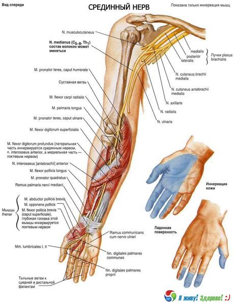
The median nerve does not give off branches on the shoulder and in the cubital fossa. On the forearm, muscle branches extend from it to the round and square pronators, the superficial flexor of the palpebrae, the long flexor of the thumb, the long palmaris muscle, the radial flexor of the carpi, and the deep flexor of the fingers (to the lateral part). The median nerve innervates all the muscles of the anterior group of the forearm, except for the medial part of the deep flexor of the fingers and the ulnar flexor of the carpi. The nerve also gives off sensory branches to the elbow joint. The largest branch of the median nerve on the forearm is the anterior interosseous nerve (n. interosseus anterior). It lies on the anterior surface of the interosseous membrane of the forearm together with the anterior interosseous artery, innervates the deep muscles of the anterior group of the forearm and the capsule of the radiocarpal joint, the interosseous membrane and the bones of the forearm.
At the level of the wrist joint, the palmar branch of the median nerve departs. It penetrates the fascia of the forearm and is directed further between the tendons of the radial flexor of the wrist and the long palmaris muscle. The palmar branch of the median nerve (r. palmaris n. mediani) innervates the skin of the lateral half of the wrist and part of the skin of the eminence of the thumb.
In the hand, the median nerve innervates the brevis muscle that abducts the pollicis; the opposing pollicis muscle; the superficial head of the brevis flexor pollicis; and the 1st and 2nd lumbrical muscles. Under the palmar aponeurosis, the median nerve divides into three common palmar digital nerves (nn. digitales palmares communes). These nerves run along the first, second, and third intermetacarpal spaces and innervate the skin of three and a half fingers on the palmar side of the hand. The first common palmar nerve innervates the 1st lumbrical muscle and gives off three cutaneous branches, the proper palmar digital nerves (nn. digitales palmares proprii). Two of them run along the radial and ulnar sides of the thumb, and the third along the radial side of the index finger. The second and third common palmar nerves give off two proper palmar digital nerves each. These nerves go to the skin of the facing sides of the first, second and third fingers and to the skin of the backs of the distal and middle phalanges of the second and third fingers. The second common palmar digital nerve also innervates the 2nd lumbrical muscle. The median nerve innervates the joints of the wrist and the first four fingers.
What do need to examine?
How to examine?