
All iLive content is medically reviewed or fact checked to ensure as much factual accuracy as possible.
We have strict sourcing guidelines and only link to reputable media sites, academic research institutions and, whenever possible, medically peer reviewed studies. Note that the numbers in parentheses ([1], [2], etc.) are clickable links to these studies.
If you feel that any of our content is inaccurate, out-of-date, or otherwise questionable, please select it and press Ctrl + Enter.
Instrumental and laboratory diagnosis of rectal cancer
Medical expert of the article

Last reviewed: 04.07.2025
Modern medicine has access to a large number of methods for the precise diagnosis of malignant bowel diseases. Such methods include both instrumental and non-instrumental examination, radiography, physiological and bacteriological tests, etc.
Diagnosis of rectal cancer is usually considered as a comprehensive examination. At the first stage, blood is taken from the patient for clinical and biochemical analysis, then feces are examined.
A digital rectal examination has also been mandatory for many years – often it is this procedure that the standard diagnostic scheme begins with. What can this or that type of examination give the doctor? Let's look at this issue in more detail.
Who to contact?
Initial stage of diagnosis of rectal cancer
If a cancerous tumor is suspected, signs such as abdominal enlargement, external fistula outlets, and enlarged lymph nodes are sometimes detected during the examination. Percussion of the abdomen can detect accumulation of fluid (ascites) or gases during intestinal perforation.
Palpation of the abdomen, despite its apparent simplicity, is considered a very valuable procedure for determining the tumor process. Thanks to palpation, it is possible to assess the degree of muscle tension, the presence of spasms and fluids, etc. The doctor must examine all lymph nodes that may be affected by the pathological process.
Examination of the perineal area allows us to see changes in the skin and anal sphincter, which can also be valuable information for diagnosis.
A digital rectal examination is considered a simple but very informative procedure. It is performed both during a preventive visit to the doctor and in the presence of complaints related to the work of the lower intestines. If such an examination is carried out carefully and competently, then it is quite possible to assess the condition of most of the rectum, and even establish a preliminary diagnosis. How the procedure is performed: the doctor inserts a finger into the rectum and feels its walls from the inside. The examination may not be very pleasant, but it is painless.
Tests for suspected rectal cancer
What lab tests are typically ordered if cancer is suspected?
- Feces for hidden blood - this method determines whether there is hidden bleeding or bleeding of the tumor. In order for the result to be as accurate as possible, feces should be submitted several times, and for prevention - once a year.
- Complete blood count – helps determine whether a person has anemia associated with hidden blood loss. Low hemoglobin levels can be one of the first indicators of cancer.
- Blood biochemistry is an assessment of carcinoembryonic antigen (CEA), the level of which determines the stage of the malignant process. Biochemistry is usually prescribed before and after surgery. As a rule, within 2 months after radical surgery, the CEA content returns to normal. If metastases are present or a relapse of oncopathology develops, its values increase again.
- A fecal DNA test is a specific analysis that is performed to detect mutated genes, that is, altered cellular structures that give rise to the development of malignant pathology.
 [ 6 ], [ 7 ], [ 8 ], [ 9 ], [ 10 ]
Tumor markers for rectal cancer
As is known, the difficulty of diagnosing a cancerous tumor is largely explained by its asymptomatic course, when the patient seeks help only when the disease has gone too far. For this reason, scientists have long been looking for a diagnostic method that would help identify the pathology as early as possible. And this method was found - this is the definition of tumor markers.
What are they? Tumor markers are unique protein substances released during the vital activity of tumor cells. They are determined in the blood or urine of a patient with oncology. At the same time, with the help of modern means, it is possible to determine an increase in the level of such substances even in the early stages of cancer.
What does the marker level indicate:
- in which organ the neoplasm may be located;
- was the prescribed treatment effective;
- is it possible for the pathology to develop again;
- is there a risk of developing cancer in the future.
There are a number of tumor markers that indicate the presence and localization of the cancer process in the rectum. These are markers such as AFP, CA 72-4, LASA-P, CA 242, CA 19-9, CYFRA 21-1.
However, there are also some disadvantages to marker analysis:
- tumor markers are not strictly specific - for example, the same indicator can indicate the presence of a process in any part of the digestive system;
- high marker levels cannot always be interpreted as the presence of a tumor;
- Some healthy people may also have these substances detected.
From all of the above, the following conclusions can be drawn: determining markers is an important procedure, but a diagnosis cannot be made based on their increase alone. Diagnostics must be taken as a whole, using all possible methods.
[ 11 ], [ 12 ], [ 13 ], [ 14 ], [ 15 ]
Instrumental diagnostics of rectal cancer
The purpose of instrumental diagnostics of the rectum is to visualize the area damaged by pathology, determine the nature of the lesion and its stage, take a tissue sample for a more detailed examination (biopsy), and also a preliminary assessment of metastasis.
- Anoscopy is a method of examining the rectum using an anoscope, an instrument that is inserted through the anal sphincter and allows the inner surface of the mucous membrane to be examined. The depth of possible examination is about 15 cm.
- Rectomanoscopy is performed using a rectoscope, which is inserted into the rectum and at a distance of up to 50 cm. This technique allows the doctor to examine the mucous membranes of the intestine, with the possibility of taking tissue samples for further analysis. The procedure cannot be called pleasant or completely painless, but as a diagnostic, it is often simply irreplaceable.
- Fibrocolonoscopy allows you to examine the inner surface of the intestine, accurately determine the location of the tumor, take pieces of material for biopsy, and even remove small polyps. With this method, you can assess the condition of the large intestine along its entire length.
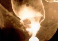
- Irrigoscopy involves the introduction of a special contrast agent into the intestinal cavity by enema, which will shade the internal cavity of the intestines when examining them on X-ray images. This procedure is used mainly in elderly patients, and also if the doctor suspects several tumor processes in the intestine at once.
- Intravenous urography may be prescribed as an additional examination method, for example, if a tumor has grown into the bladder.
- Ultrasound monitoring of abdominal organs is used to detect metastases. If the procedure is performed with fluid accumulation (ascites), then ultrasound allows you to estimate its volume.
- The computed tomography method is used to determine the growth of a tumor into nearby organs, find metastases, and check the nearest lymph nodes.
- Diagnostic laparoscopy is a minimally invasive surgical procedure where the abdominal wall is punctured in several places and a special camera is inserted through the punctures, which allows not only to see the presence of metastases in the abdominal cavity, but also to remove small objects.
I would like to dwell separately on the magnetic resonance imaging method, which, although not considered a priority examination, can sometimes provide very important information to the doctor. Determining the extent of tumor growth, selecting a therapeutic regimen, assessing the need and scope of surgery - these are exactly the cases when this procedure is simply necessary. In addition, MRI allows you to monitor and evaluate the course of cancer treatment and determine further tactics in relation to the patient.
Rectal cancer on MRI will be better visualized and assessed by the doctor if you follow these recommendations:
- Before the procedure, you should cleanse the rectum - this can be done with a laxative or a regular cleansing enema;
- 1-1.5 hours before the procedure, you must empty your bladder, after which drinking is not allowed until the end of the examination;
- It is recommended to take 3 tablets of drotaverine (No-shpa) approximately one hour before the MRI.
If everything is done correctly, the doctor will be able to easily perform the following actions:
- see the tumor itself, including its borders;
- determine the relationship of the neoplasm to the pelvic organs and sphincter;
- determine the condition of the pelvic muscles;
- assess the condition of the lymphatic system in the pelvis;
- monitor the effectiveness of surgery, chemotherapy or radiation, and track the disease over time.
Histological and cytological studies
In order to distinguish a benign disease from a malignant one, a study such as a biopsy is used, followed by a histological analysis. Thanks to a biopsy, it is possible to state with great accuracy the presence or absence of a cancerous tumor in the tissues being examined. The diagnostic method involves the removal of a small element of tumor tissue - the entire process is carried out during a rectoscopy and does not cause any additional discomfort to the patient. The obtained piece of tissue is subjected to histological and cytological assessment.
In addition to rectoscopy, the doctor can take the material he needs during laparoscopy, surgery or fibrocolonoscopy. •
Histological examination involves examining a sample of removed tissue using a microscopic method and can be carried out on an urgent or planned basis:
- urgent histology is performed in about half an hour, when a quick result is needed. The sample is first frozen, then treated with specific dyes and examined using a microscope;
- planned histology usually lasts at least 5 days. The obtained sample is covered with a special liquid and paraffin, and painted. This method of research is considered more complicated if compared with urgent histology. However, its results are more accurate and reliable.
As a rule, in order to ensure that the results of a histological examination are not questioned in the future, it is carried out by at least two specialists. •
Cytological examination is an assessment of the cellular structures of tissue, which allows one to notice malignant changes in them. How does this analysis differ from histological? In that the cytological method involves examining not a tissue section, but individual tumor cells.
The following biological material can be used to perform cytology:
- tissue samples taken during biopsy from the required section of the intestine;
- purulent or mucous discharge from the intestinal cavity;
- samples of mucous tissue prints from the required section of the intestine.
Only the above methods allow us to determine with certainty which tumor needs to be treated: benign or malignant.
Differential diagnosis of rectal cancer
Rectal cancer must be distinguished from the following diseases:
- benign polyps;
- chronic ulcerative proctosigmoiditis of dysenteric, amoebic and tuberculous origin;
- colitis, rectal prolapse, non-specific granuloma;
- syphilis, actinomycosis;
- melanoblastomas of the anal rectum;
- tumor growth from the uterus, vagina, prostate;
- malignant carcinoid;
- hemorrhoids and anal sphincter fissures.
In order to differentiate a cancerous tumor in the rectum from polyps, an endoscopic examination is prescribed - colonoscopy. This method allows you to see in the lumen of the intestine not only large polyps, but also ulcers of the mucous membrane, inflammatory elements, small flattened polyps, deformed vessels, etc. The same method can be used to differentiate cancer and proctosigmoiditis - an inflammatory process in the sigmoid and rectum.
[ 16 ], [ 17 ], [ 18 ], [ 19 ], [ 20 ], [ 21 ], [ 22 ]
How to distinguish hemorrhoids from rectal cancer?
Sometimes certain signs play a decisive role, by which one can distinguish a tumor process from ordinary hemorrhoids.
- If the patient has previously been diagnosed with polyps, there is a risk that they will degenerate into a cancerous tumor.
- Hemorrhoidal blood is released at the end of the act of defecation, in the form of stripes and spots on top of the feces. In the case of cancer, the blood is not on the surface, but mixed with feces.
- In case of a tumor, there may be mucous discharge before defecation, sometimes with pus and pieces of tissue.
- With an extensive tumor, the feces are ribbon-like, and difficult defecation can last for a long time, up to several days.
- The tumor process is accompanied by emaciation of patients, weakness and lethargy.
- In the presence of metastases, the functioning of other organs begins to deteriorate.
However, in any case, to establish an accurate diagnosis, it is necessary to conduct a biopsy with a histological examination of the tissues. Only after this can one confidently state the presence or absence of a malignant process.
Rectal cancer diagnostics should be performed at the slightest suspicion of its presence. It is very important to detect a malignant disease as early as possible - this will allow timely treatment, which will significantly improve the prognosis and speed up recovery.