
All iLive content is medically reviewed or fact checked to ensure as much factual accuracy as possible.
We have strict sourcing guidelines and only link to reputable media sites, academic research institutions and, whenever possible, medically peer reviewed studies. Note that the numbers in parentheses ([1], [2], etc.) are clickable links to these studies.
If you feel that any of our content is inaccurate, out-of-date, or otherwise questionable, please select it and press Ctrl + Enter.
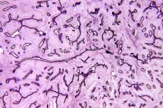
Histology of breast tissue
Medical expert of the article

Last reviewed: 04.07.2025
When the results of mammography or ultrasound of the mammary gland show changes that may be of an oncological nature, a tissue sample of the pathological formation is taken - a biopsy is done. The resulting sample is studied by pathomorphologists and its cellular structure is determined, that is, a histology of the mammary gland tissue is performed. Histology, as the most important scientific tool of biology and medicine, is the only way to identify abnormal tumor cells.
Therefore, histology of breast tumors serves as the most accurate diagnostic method in oncology and helps to choose the most effective treatment path.
Indications for histology of breast tissue
The main indications for biopsy and histology of breast tissue include the possibility of a malignant nature of the pathology in the case of:
- intense focal or diffuse hyperplasia in various tissues and structures of the mammary glands;
- fibroadenosis, including phyllodes fibroadenoma;
- cystic lesions of the mammary glands;
- intraductal papillomatosis;
- tissue necrosis (glandular, fibrous, fatty);
- breast cancer, its recurrences and metastases.
The most important signs that give grounds to suspect the malignancy of the pathological process in the tissues of the mammary glands and resort to their examination at the cellular (cytological) level are manifested in the form of intra-tissue seals (both palpable and recorded on a mammogram or ultrasound); abnormal discharge from the nipples; deformations, discoloration or ulceration of the nipple-areolar zone of the breast; various changes in the skin of the mammary gland; an increase in the size of regional lymph nodes, etc.
For more information on biopsy methods and procedures, see the publication Breast Biopsy.
Who to contact?
Decoding the histology of the mammary gland: main indicators
The study of the morphology and biochemical characteristics of the mammary gland tissue is carried out on the thinnest sections under a light or electron microscope. Special histological dyes are used to improve visualization and more accurate analysis of tissue structures. Thanks to phase-contrast, fluorescent, interference and other microscopy methods, as well as the study of the chemical composition of cells by cytospectrophotometry, the results of histological studies and decoding of the histology of the mammary gland make it possible to carry out differential diagnostics of tumors - benign and cancerous.
Breast cancer histology can determine:
- morphological type of tumor and its histogenesis;
- degree of malignancy (malignancy);
- hormonal status of the neoplasm;
- degree of distribution.
Depending on the shape of the tumor cells, specialists distinguish histological forms of malignant neoplasms. If the cells are similar to brain tissue, then medullary cancer is determined; if the cells are tubular, tubular cancer is determined; with a high content of mucin, mucous.
Deciphering the histology of the mammary gland by the degree of malignancy or, as specialists define it, the degree of tumor differentiation, is based on studying the structure of neoplasm cells with the isolation of mutated cells (cell anaplasia) and determining their percentage in relation to healthy cells. The lowest degree (grade) of malignancy is the first (GI), the highest is GIV.
The histology of fibroadenoma of the mammary gland - if the pathology is benign - should have a GX degree according to this indicator, which means that “the degree of malignancy cannot be assessed” (i.e., oncology is not detected).
The immunohistochemical method determines the substances that make up the cells, and the immunocytochemical method determines tissue biomarkers of membrane receptors of estrogen (ER) and progesterone (PR) and epidermal growth factor (HER2/neu). Immunofluorescence automated quantitative analysis (AQUA) determines the proliferative activity of the tumor (Ki 67), i.e. the intensity of its cell mitosis.
Decoding the histology of the mammary gland in relation to markers of steroid hormones: positive results for estrogen receptors (ER+) and progesterone (PR+) indicate that the growth of cancer cells depends on these hormones. According to oncologists-mammologists, histology in adenocarcinoma of the mammary gland (glandular cancer or ductal carcinoma) with such results is noted in 75-80% of cases of the disease in 40-45-year-old women. And hormone-negative cancer (ER- and PR-) is diagnosed in patients after 50-55 years. The presence of these receptors helps to determine both the degree of risk of relapse and the optimal hormonal treatment of the tumor.
When histology results show increased activity of the human epidermal growth factor receptor HER2 on the membranes of cancer cells, the so-called HER2-positive cancer is defined, and such cancer is characterized by rapid development.
If the histology results are ER-, PR-, and HER2-, the tumor is called triple negative. Triple negative cancers account for about 15% of invasive breast cancers and are the most common type diagnosed in women with a BRCA1 gene mutation.
A high level of Ki-67 is defined in the range of 15-25%, more than 40% is considered a very high indicator and from a prognostic point of view means an unfavorable outcome of breast cancer. In addition, the marker of tumor proliferative activity provides information on the effectiveness of preoperative systemic therapy - when comparing its level before and after surgery.
Histology of breast tissue is the most important method for examining the condition of the breast and determining the nature of its pathologies.
 [ 6 ]
[ 6 ]