
All iLive content is medically reviewed or fact checked to ensure as much factual accuracy as possible.
We have strict sourcing guidelines and only link to reputable media sites, academic research institutions and, whenever possible, medically peer reviewed studies. Note that the numbers in parentheses ([1], [2], etc.) are clickable links to these studies.
If you feel that any of our content is inaccurate, out-of-date, or otherwise questionable, please select it and press Ctrl + Enter.
Examination of comatose patients
Medical expert of the article

Last reviewed: 07.07.2025
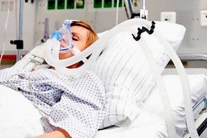
Coma is the deepest depression of consciousness, in which the patient is unable to make speech contact, follow commands, open eyes, and respond to painful stimuli in a coordinated manner. Coma develops with bilateral diffuse damage (anatomical or metabolic) to the cortex and subcortex of the cerebral hemispheres, the brain stem, or with combined damage at these levels.
General principles of examination
When examining patients in a comatose state, it is advisable to adhere to the following steps.
- Evaluation of vital functions - respiration and circulation. Determine the patency of the airways, the nature of breathing, the presence of pathological types of breathing; the frequency, volume and rhythm of the pulse; arterial pressure.
- Assessment of the degree of depression of consciousness (depth of coma).
- A brief explanation of the circumstances of the development of a coma, the factors preceding it, and the rate of loss of consciousness.
- General examination of the patient, during which special attention should be paid to signs of injury (abrasions, bruises, swelling, etc.); bleeding from the ears and nose; the presence of a periorbital hematoma; changes in skin color, moisture, temperature; bad breath; body temperature; any other symptoms of acute pathology.
- A brief neurological examination, with particular attention paid to brainstem reflexes ( pupil reactions, position and movements of the eyeballs); posture, muscle tone, deep reflexes, pathological signs, involuntary motor activity; symptoms of irritation of the meninges.
Examination of a patient in a comatose state must be combined with emergency measures to eliminate life-threatening respiratory and circulatory disorders.
Vital function assessment
Vital functions include, first of all, breathing and blood circulation. The patency of the airways, breathing characteristics, pulse and blood pressure are assessed. The results of such an assessment are extremely important for timely correction of the detected disorders.
Pathological types of breathing are often detected in patients in coma. The type of breathing disorder can be used to suggest the localization and sometimes the nature of the pathological process.
- Cheyne-Stokes respiration is a series of gradually increasing and then decreasing in frequency and depth of breaths, alternating with periods of shallow breathing or short pauses in breathing (the amplitude and frequency of respiratory movements increase and decrease in a wave-like manner until a pause in respiratory movements appears). Periods of hyperpnea are longer than periods of apnea. Cheyne-Stokes respiration indicates damage to the hypothalamic (diencephalic) region or bilateral dysfunction of the cerebral hemispheres. It is observed in metabolic disorders, rapid increase in intracranial pressure, somatic diseases (for example, in severe heart failure).
- Shallow, slow, but rhythmic breathing is characteristic of comas that develop against the background of metabolic disorders or the toxic effects of drugs.
- Kussmaul breathing is a deep and noisy breathing, characterized by rhythmic rare respiratory cycles, deep noisy inspiration and forced expiration. It is typical for ketoacidotic, hepatic, uremic coma and other conditions accompanied by metabolic acidosis ( lactic acidosis, organic acid poisoning). Hyperventilation can also occur with respiratory alkalosis ( hepatic encephalopathy, salicylate poisoning) or hypoxemia.
- True central neurogenic hyperventilation ("machine breathing") is rapid (over 30 per minute) rhythmic deep breathing, usually with a reduced amplitude of chest excursion; it occurs with dysfunction of the pons or midbrain and usually serves as an unfavorable prognostic sign, since it indicates a deepening of the coma. The neurogenic nature of hyperventilation is established only after excluding its other possible causes, which were mentioned above.
- Apneustic breathing is characterized by an extended inhalation followed by holding the breath at the height of inhalation (“inspiratory spasm”) and has a topical significance, indicating a focus in the area of the pons (for example, with occlusion of the basilar artery).
- Cluster breathing: periods of rapid, uneven breathing alternate with periods of apnea; may resemble Cheyne-Stokes breathing, combined with various types of difficulty breathing. Occurs with damage to the upper parts of the medulla oblongata or lower parts of the pons and serves as a threatening sign. One of the options is Biot's breathing: frequent, even breathing movements separated by periods of apnea. It is characteristic of damage to the pons.
- Ataxic breathing, characterized by an arrhythmic alternation of deep and shallow breaths with pauses, occurs when the medulla oblongata (respiratory center) is damaged. In this case, the sensitivity of cerebral structures to sedatives and other drugs increases, an increase in the dose of which easily causes respiratory arrest. Such breathing is usually preterminal.
- Agonal sighs are single, rare, short and deep convulsive respiratory movements against the background of apnea; they occur during agony and usually precede a complete cessation of breathing.
 [
[ Blood pressure and pulse
A decrease in blood pressure may occur not only as a result of pathological conditions that lead to coma (internal bleeding, myocardial infarction ), but also as a result of suppression of the medulla oblongata function (alcohol and barbiturate poisoning). Arterial hypertension may also either reflect the process that led to coma or be a consequence of dysfunction of the brainstem structures. Thus, an increase in intracranial pressure leads to an increase in systolic and diastolic blood pressure, while the pulse is usually slow. The combination of arterial hypertension with bradycardia (Cushing's phenomenon) indicates an increase in intracranial pressure.
Estimating the depth of coma
The most well-known rapid quantitative method for determining the depth of coma is the use of the Glasgow Coma Scale. According to this approach, the severity of depression of consciousness is determined based on the assessment of the patient's reactions: opening of the eyes, speech reaction, motor reaction to pain. The total score on the Glasgow Coma Scale can range from 3 to 15 points. A score of 8 points or less indicates the presence of a coma. The use of this scale allows only a preliminary assessment of the depth of the disorder of consciousness; a more accurate conclusion is made after a neurological examination.
- Mild (grade I) coma is characterized by the development of general motor restlessness or withdrawal of a limb in response to a painful stimulus, a reflex response in the form of sneezing when the mucous membrane of the nose is irritated with cotton wool soaked in ammonia; facial reactions on the same side when percussing the zygomatic arch. Corneal reflexes and pupillary reactions to light are preserved, swallowing is not impaired, breathing and blood circulation are sufficient to maintain the body's vital functions. Urination is involuntary; urinary retention is possible.
- Severe (grade II) coma is characterized by a complete absence of motor response to sound and moderate pain stimuli and the occurrence of protective reflexes to strong pain stimuli. Pathological types of breathing, arterial hypotension and cardiac arrhythmias are observed. The pupils are often narrow, less often wide, their reactions to light and corneal reflexes are weakened. Swallowing is impaired, but when liquid enters the respiratory tract, coughing movements occur, indicating partial preservation of bulbar functions. Deep reflexes are suppressed. The grasping and proboscis reflexes, Babinski's symptom are revealed.
- Deep (grade III) coma is characterized by the extinction of all reflex acts, including vital ones. Typical are inadequate breathing (bradypnea with a frequency of less than 10 per minute, etc.), weakness of cardiac activity (collapse, arrhythmia, cyanosis of the skin and mucous membranes), absence of motor reactions, muscular hypotonia. The eyeballs are in a neutral position, the pupils are wide, their reaction to light and corneal reflexes are absent, swallowing is impaired.
Clarification of the circumstances of the development of coma
Information about the circumstances of the coma development, the rate of loss of consciousness, and the illnesses the patient suffered from are obtained from the patient's relatives or people around him. This information is important for establishing the cause of the coma.
- A history of stroke, arterial hypertension, vasculitis or heart disease (may indicate a vascular nature of the coma).
- In a patient with diabetes mellitus, coma may be a consequence of diabetic ketoacidosis (ketoacidotic coma), hyperosmolar nonketogenic state (hyperosmolar coma), lactic acidosis (hyperlactacidemic coma), insulin-induced hypoglycemia (hypoglycemic coma).
- Coma in a patient with epilepsy may result from status epilepticus or from traumatic brain injury sustained during a seizure.
- A recent history of head trauma suggests causes of coma such as brain contusion, intracerebral hematoma, and diffuse axonal injury.
- A history of alcoholism increases the likelihood of alcoholic coma, hepatic coma, Wernicke encephalopathy, and also allows one to suspect head trauma as one of the possible causes of coma.
- Coma can be a consequence of an overdose of insulin, sedatives and sleeping pills, antidepressants, neuroleptics, narcotics, barbiturates.
- In infections, both metabolic ( meningitis, encephalitis, sepsis, neurosarcoidosis) and structural ( herpes encephalitis, brain abscess with the development of dislocation syndrome) causes of coma are possible.
General examination of the patient
Examination of the skin and mucous membranes, as well as examination of the chest, abdomen and extremities, carried out according to general rules, are aimed at identifying manifestations specific to certain comas.
- It is necessary to carefully examine the patient for signs of injury (bleeding, bruising, hematomas, tissue swelling). Thus, signs of a basal skull fracture may include Battle's symptom (hematoma in the mastoid process area), local pain, hemorrhage into the conjunctiva and periorbital tissue ("glasses"), bleeding and cerebrospinal fluid rhinorrhea from the ear and nose.
- When assessing the condition of the skin, the following are of differential diagnostic importance: "spiders", abrasions, venous pattern, injection marks; skin turgor, dryness or moisture. Pink or scarlet skin is characteristic of carbon monoxide and cyanide poisoning, icteric skin - of liver disease, yellowish-ash skin with a whitish tint on the lips - of uremia, severe pallor - of anemia and internal bleeding, cyanotic skin with a slate-gray or black-blue tint - of poisoning with methemoglobin-forming poisons, brown skin - of bromide poisoning.
- Information about the condition of the sclera, the tone of the eyeballs, body temperature, and the color of the vomit is important.
- The density of the eyeballs is determined by pressing the eyelids with the flesh of the nail phalanx of the index finger. A decrease in skin turgor and density of the eyeballs is found in uremia, chlorpenia, food toxicoinfection, alimentary dystrophy, hyperglycemia, dehydration of the body of any genesis. On the contrary, in those who have received a severe craniocerebral injury, even with a sharp decrease in hemodynamic parameters, the density of the eyeballs is increased, and the possibility of their displacement into the depth of the orbit is limited. Injection of the sclera is most often observed in subarachnoid hemorrhage, epilepsy, fat embolism of cerebral vessels, alcohol intoxication.
- Multiple whitish scars on the lateral surfaces of the tongue with fresh bites are formed as a result of repeated convulsive seizures.
- Hyperthermia is observed in meningitis, encephalitis, septic thrombosis of the cerebral sinuses, thyrotoxicosis, food poisoning, pneumonia, dehydration, poisoning with atropine-like drugs and tricyclic antidepressants, intracranial hematomas with symptoms of damage to the brainstem and hypothalamus. Hypothermia is characteristic of chlorpenia, uremia, alimentary exhaustion, adrenal insufficiency, as well as poisoning with barbiturates and tranquilizers.
Assessment of neurological status
Neurological examination is aimed at assessing general motor reactions, brainstem reflexes and identifying symptoms of irritation of the meninges.
Motor sphere
We evaluate the patient’s posture, muscle tone and deep reflexes, spontaneous and provoked motor activity.
Pathological postures:
- If the patient lies in a natural position, as in normal sleep, one can think of a shallow coma, which is confirmed by the preservation of yawning and sneezing. Other reflex acts in the form of coughing, swallowing or hiccups are preserved even with a deeper depression of consciousness.
- Pathological postures, mainly flexor or extensor, are sometimes observed in a patient in a coma. Sometimes terms borrowed from pathophysiology are used, such as "decorticate" and "decerebrate rigidity". In decorticate rigidity, the arms are brought to the body, bent at the elbows and wrists, the hands are supinated; the legs are extended at the hips and knees, rotated inward, the feet are in a position of plantar flexion. This posture develops due to the loss of inhibitory corticospinal influences and indicates a lesion above the midbrain. In decerebrate rigidity, the head is thrown back (opisthotonus), the teeth are clenched, the arms are extended and rotated inward, the fingers are bent, the legs are straightened and rotated inward, the feet are in a position of plantar flexion. Pinching of the skin on the trunk and extremities causes protective spinal reflexes, which in the legs often take the form of triple flexion (in the hip, knee and ankle joints). Decerebrate rigidity indicates damage to the upper part of the brainstem at the level between the red and vestibular nuclei with the loss of central inhibitory effects on peripheral motor neurons with disinhibition of descending vestibular tonic impulses. The decorticate posture, compared with the decerebrate posture, indicates a more rostral localization of the lesion and a more favorable prognosis; however, it is impossible to reliably judge the localization of the lesion based on the patient's posture alone.
- Asymmetry of limb position and unusual posture of individual body parts may be of diagnostic value. Thus, in a patient with hemiplegia, which developed as a result of damage to the internal capsule and nodes of the base of the brain, muscle tone in the affected limbs is reduced in the acute period of the disease. If such a patient is in a coma, then his foot on the side of paralysis is rotated outward (Bogolepov's symptom). Fixed deviation of the head backwards and to the side is often observed in patients with tumors of the posterior cranial fossa. A posture with a thrown back head and a curved back is often a sign of irritation of the meninges (with subarachnoid hemorrhage, meningitis). Bringing the legs to the stomach is observed in many patients with uremic coma.
Muscle tone and spontaneous motor activity
- Repetitive twitching of the facial muscles, fingers, and/or feet may be the only manifestation of an epileptic seizure. Full-blown epileptic seizures have no topical diagnostic significance, but they indicate the preservation of the corticomuscular pathway.
- Multifocal myoclonic seizures are often a sign of metabolic brain damage (azotemia, drug poisoning) or late-stage Creutzfeldt-Jakob disease. Asterixis also indicates metabolic encephalopathy (in uremia, liver failure).
- The preservation of complex reflex actions, such as defensive movements and other purposeful actions (such as scratching the nose in response to tickling the nostril), indicates the preservation of the pyramidal system on the corresponding side. The absence of automated movements in some limbs in a comatose patient indicates paralysis of that side.
- Hormetonic convulsions (attacks of increased muscle tone, usually in paralyzed limbs and following each other with short pauses) are observed with hemorrhage into the cerebral ventricles. The duration of such tonic spasms varies from a few seconds to several minutes. As a rule, paroxysmal increase in tone in the arm involves the adductor muscles of the shoulder and the pronators of the forearm, and in the legs - the adductor muscles of the thigh and the extensors of the shin.
Initiated motor activity - movements that occur reflexively in response to external stimulation (pricks, pinches, stroking).
- When a painful stimulus causes a targeted abduction of a limb without its pronounced flexion, one can think about the preservation of the cortical-muscular pathway to this limb. If a similar targeted abduction occurs in all limbs during their painful stimulation, then the patient's motor disorders are minimal. Thus, abduction of a limb is a sign of relative preservation of the motor system. On the contrary, if in response to irritation of the limbs a patient in a coma takes stereotypical poses, this indicates severe bilateral damage to the pyramidal systems.
- The detection of a grasping reflex upon stimulation of the palmar surface of the hand indicates damage to the opposite frontal lobe.
- The phenomenon of counter-continence with the emergence of resistance to passive movements of the limbs is characteristic of diffuse damage to the anterior parts of the brain due to a metabolic, vascular or atrophic pathological process.
- Normal muscle tone and preservation of deep reflexes indicate the intactness of the cortex and corticospinal tract. Asymmetry of muscle tone and reflexes is observed with supratentorial localization of the lesion; it is not characteristic of metabolic coma. Symmetrical decrease in muscle tone and suppression of deep reflexes are typical of metabolic coma. Changing muscle tone and reflexes are usually observed in epileptic seizures and psychiatric pathology.
Brainstem reflexes play an important role in assessing cerebral coma and reflect the degree of preservation of the cranial nerve nuclei (while deep reflexes in the limbs are spinal reflexes, so their diagnostic value in patients in a coma is limited). Impaired brainstem reflexes most likely indicate that the depression of consciousness is associated with dysfunction of the ascending activating system of the reticular formation of the brainstem. Conversely, the preservation of brainstem reflexes indicates the intactness of the brainstem structures (coma is most likely associated with extensive bilateral damage to the cerebral hemispheres). To assess brainstem function, pupillary reactions, corneal reflex, and eye movements are primarily examined.
- The size and shape of the pupils, their direct and consensual reactions to light are assessed.
- Unilateral pupil dilation with no reaction to light in a patient in a coma (Hutchinson's pupil) most often indicates compression of the oculomotor nerve as a result of temporotentorial herniation, especially if the pupil dilation is combined with downward and outward deviation of the eyeball. Less often, a dilated pupil that does not react to light is observed in cases of damage or compression of the midbrain itself.
- Bilateral pinpoint pupils with a weak reaction to light (in this case, a magnifying glass is used to assess pupillary reactions) indicate damage to the pontine tegmentum with descending sympathetic pathways passing through this area (sympathetic innervation of the pupils is lost and parasympathetic innervation begins to predominate, since the Edinger-Westphal nuclei remain intact).
- Bilateral fixed mydriasis (wide areactive pupils with a diameter of 4-6 mm) is observed in severe damage to the midbrain with destruction of the parasympathetic nuclei of the oculomotor nerve, as well as in botulism and poisoning with atropine, cocaine, and mushrooms.
- The pupillary response to light can serve as a clue in determining the cause of coma. In metabolic disorders, pupillary response to light in a comatose patient most often persists for a long time, even in the absence of all other neurological reactions (except for hypoxic encephalopathy and poisoning with anticholinergic drugs), while in focal brain lesions they disappear early. For example, in patients with craniocerebral trauma, a weakened pupillary response to light is almost always observed and does not indicate a poor prognosis.
- Preservation of pupillary responses is a sign of the integrity of the midbrain. Equal pupils that react to light indicate a toxic/metabolic coma, with some exceptions. Metabolic causes of fixed mydriasis include hypoxic encephalopathy and poisoning with anticholinergics (atropine) or botulinum toxin. Drug poisoning, as well as the use of narcotic analgesics or pilocarpine, cause constriction of the pupils (miosis) with a weak reaction to light, which can sometimes only be detected with a magnifying glass.
- Attention is paid to the closure of the eyelids (i.e., to the preservation of the connections between the V and VII pairs of cranial nerves) and to the symmetry of the corneal reflexes. Corneal reflexes are characterized by a different pattern than pupillary reactions to light: in case of poisoning with drugs that depress the central nervous system, the corneal reflex decreases or disappears quite early, while in coma caused by traumatic brain injury, on the contrary, the disappearance of the corneal reflex indicates the severity of the injury and is an unfavorable prognostic sign. Thus, the preservation of pupillary reactions in a patient in deep coma in the absence of corneal reflexes and eye movements allows us to suspect a metabolic disorder (for example, hypoglycemia ) or poisoning with drugs (in particular, barbiturates).
- Evaluation of the position and movements of the eyeballs. When the eyelids of a patient in a coma are lifted, they slowly fall. If the eyelids do not close completely on one side, it is possible to assume damage to the facial nerve (nuclear damage on this side or supranuclear on the opposite side). If the patient is not in a coma, but in a hysterical seizure, then resistance is experienced when the eyes are passively opened. The preservation of blinking in a patient in a coma indicates the functioning of the reticular formation of the pons. After opening the eyelids, the position of the eyeballs and spontaneous eye movements are assessed. In healthy people, the axes of the eyeballs are parallel in the waking state, and in a drowsy state, a deviation of the eyeballs occurs. In patients in a coma, the eyeballs may occupy a position along the midline, be diverged along the horizontal or vertical axis, or be diverted up/down or to the side.
- Persistent consensual deviation of the eyeballs to the side may indicate damage to the ipsilateral hemisphere or contralateral area of the pons. When the frontal lobe of the cerebral hemisphere is damaged (the frontal center of horizontal gaze), the eyeballs "look" towards the lesion, "turning away" from the paralyzed limbs. Reflex movements of the eyeballs are preserved (i.e., deviation of the eyeballs in case of damage to the frontal lobe can be overcome by turning the head sharply - the "doll's eyes" phenomenon is preserved). When the horizontal gaze center in the pons tegmentum is damaged, the eyes, on the contrary, "turn away" from the lesion and "look" at the paralyzed limbs. It is not possible to overcome the deviation of the eyeballs by turning the head due to the suppression of the vestibulo-ocular reflex (the "doll's eyes" phenomenon is absent). There is only one exception to the rule that supratentorial lesions cause the eyes to deviate toward the site of destruction: with hemorrhage in the medial parts of the thalamus, an “incorrect” deviation of gaze may occur - the eyes “turn away” from the affected thalamus and “look” at the paralyzed limbs.
- Deviation of the eyeballs downwards in combination with a violation of their convergence is observed in cases of damage to the thalamus or pretectal region of the midbrain. May be combined with pupillary unresponsiveness (Parinaud's syndrome). Usually occurs in metabolic coma (especially in cases of barbiturate poisoning).
- Divergence of the eyeballs along the vertical or horizontal axis, or the upward/downward or lateral deviation of both eyeballs, usually indicates a focal lesion of the brain.
- Inward deviation of one eyeball occurs with paralysis of the lateral rectus muscle of the eye and indicates damage to the abducens nerve (most likely in the region of its nucleus in the pons). Inward deviation of both eyeballs develops as a result of bilateral damage to the abducens nerves as a symptom during intracranial hypertension. Outward deviation of one eyeball indicates damage to the medial rectus muscle of the eye with insufficiency of the function of the nucleus of the oculomotor nerve.
- Vertical divergence of the eyeballs with a downward and inward deviation of the eyeball on the affected side, and upward and outward on the opposite side (Hertwig-Magendie symptom) is characteristic of a violation of the vestibular connections with the medial longitudinal fasciculus. This symptom is observed in tumors of the posterior cranial fossa or in circulatory disorders in the area of the brain stem and cerebellum, as well as in the localization of the tumor in the cerebellar hemispheres with pressure on the roof of the midbrain.
- Constant tonic downward deviation of the eyeballs (the setting sun phenomenon) most often occurs with hydrocephalus with dilation of the third ventricle.
- Spontaneous eye movements. "Floating" eye movements in the horizontal direction are sometimes observed in mild coma; they are of no particular importance for topical diagnostics. Their appearance indicates the preservation of the brainstem structures (nuclei of the third pair of cranial nerves and the medial longitudinal fasciculus). Normal nystagmus is not typical for patients in coma, since in coma the interaction between the cochleovestibular apparatus of the brainstem (formation of the slow phase of nystagmus) and the cerebral hemispheres (formation of the fast phase of nystagmus), which is necessary for its development, is disrupted, and voluntary fixation of the gaze is absent.
- Reflex movements of the eyeballs (oculocephalic or vestibulo-ocular reflex) are mediated by pathways that go through the brainstem, so inhibition of these reactions indicates damage to the brainstem structures. Reflex movements of the eyeballs cause the "doll's eye" test and, less often, the cold test (introduction of cold water into the external auditory canal).
Meningeal signs (particularly nuchal rigidity) may be a sign of meningitis, brain injury, or subarachnoid hemorrhage. They should not be tested if a cervical spine fracture is suspected.
What do need to examine?