
All iLive content is medically reviewed or fact checked to ensure as much factual accuracy as possible.
We have strict sourcing guidelines and only link to reputable media sites, academic research institutions and, whenever possible, medically peer reviewed studies. Note that the numbers in parentheses ([1], [2], etc.) are clickable links to these studies.
If you feel that any of our content is inaccurate, out-of-date, or otherwise questionable, please select it and press Ctrl + Enter.
The muscle that straightens the spine.
Medical expert of the article

Last reviewed: 07.07.2025
The erector spinae muscle is the strongest of the autochthonous muscles of the back, extending along the entire length of the spine - from the sacrum to the base of the skull. It lies in front of the trapezius, rhomboid, posterior serratus muscles, and the latissimus dorsi. Behind, the erector spinae muscle is covered by the superficial layer of the thoracolumbar fascia. The muscle begins with thick tendinous bundles on the dorsal surface of the sacrum, spinous processes, supraspinous ligaments of the lumbar, 12th and 11th thoracic vertebrae, posterior segment of the ilium crest, and thoracolumbar fascia. Some of the tendinous bundles that begin in the sacrum merge with the bundles of the sacrotuberous and dorsal sacroiliac ligaments.
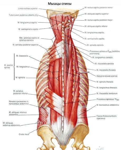
At the level of the upper lumbar vertebrae, the erector spinae muscle is divided into three tracts: lateral, intermediate, and medial. Each tract is given its own name. The lateral tract is the iliocostalis muscle, the intermediate tract is the longissimus muscle, and the medial tract is the spinalis muscle. Each of these muscles is further subdivided into parts.
The structural features of the erector spinae muscle developed during anthropogenesis in connection with upright walking. The fact that the muscle is highly developed and has a common origin on the pelvic bones, and is further divided into separate tracts that are widely attached to the vertebrae, ribs and the base of the skull, can be explained by the fact that it performs a vital function - it holds the body in an upright position. At the same time, the division of the muscle into separate tracts, the subdivision of the latter at different levels of the dorsal side of the body into shorter muscles that have a shorter length between the origin and attachment points, allows the muscle to act selectively. For example, when the iliocostalis lumborum muscle contracts, the corresponding ribs are pulled downwards, thereby creating support for the manifestation of the force of the diaphragm when it contracts, etc.
The function of the entire erector spinae muscle is fairly accurately reflected by its name. Since the muscle's components originate on the vertebrae, it can act with its entire mass as an extensor of the spine (trunk) and head, overcoming the resistance of the ventral muscles and the body's gravity. By contracting with separate parts on both sides, this muscle can lower the ribs, straighten various sections of the spine, and throw the head back. With a unilateral contraction, it tilts the spine (trunk) to the same side. The muscle develops greater strength when it prevents the body from falling forward under the action of ventrally located muscles that have a greater lever of action on the spinal column.
What do need to examine?
How to examine?