
All iLive content is medically reviewed or fact checked to ensure as much factual accuracy as possible.
We have strict sourcing guidelines and only link to reputable media sites, academic research institutions and, whenever possible, medically peer reviewed studies. Note that the numbers in parentheses ([1], [2], etc.) are clickable links to these studies.
If you feel that any of our content is inaccurate, out-of-date, or otherwise questionable, please select it and press Ctrl + Enter.
Peculiarities of ECG in children
Medical expert of the article

Last reviewed: 03.07.2025
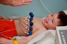
ECG in children is important for diagnosing heart disease. The technique of taking an ECG, the lead system and the theoretical basis of the method are common to all age groups. However, the interpretation of ECG results in children is more complex due to age differences in individual ECG indicators.
 [ 1 ], [ 2 ], [ 3 ], [ 4 ], [ 5 ], [ 6 ], [ 7 ], [ 8 ], [ 9 ]
ECG waves and intervals in children
The P wave reflects the spread of excitation in the atrial myocardium. The first half of the wave to its apex corresponds to excitation of the right atrium, the second - to the left. The duration of the P wave in healthy children does not exceed 0.1 s. In the III standard lead, the wave can be negative, biphasic or smoothed.
The P-Q or P-R interval includes the P wave and the isoelectric line from P to the Q or R wave. The interval changes with the pulse rate, and its normal values are estimated from tables.
P-Q interval and QRS complex in children (duration in seconds in lead II), according to Yu. M. Belozerov
Age, |
R-Q |
QRS |
||||
10 |
50 |
90 |
10 |
50 |
90 |
|
1 |
0.08 |
0.10 |
0.13 |
0.053 |
0.065 |
0,077 |
2 |
0.08 |
0.11 |
0.14 |
0.053 |
0.065 |
0,077 |
3 |
0.08 |
0.11 |
0.14 |
0.053 |
0.064 |
0,077 |
4 |
0.08 |
0.12 |
0.14 |
0.063 |
0.072 |
0.082 |
5 |
0.09 |
0.12 |
0.14 |
0.063 |
0,070 |
0.083 |
6 |
0.09 |
0.12 |
0.15 |
0.053 |
0.068 |
0,079 |
7 |
0.10 |
0.12 |
0.15 |
0.062 |
0.067 |
0.081 |
8 |
0.10 |
0.13 |
0.16 |
0.053 |
0.067 |
0.081 |
9 |
0.10 |
0.13 |
0.17 |
0.053 |
0.073 |
0.085 |
10 |
0.11 |
0.14 |
0.17 |
0.053 |
0.072 |
0.086 |
11 |
0.11 |
0.14 |
0.16 |
0.053 |
0.073 |
0.085 |
12 |
0.11 |
0.14 |
0.16 |
0.053 |
0.073 |
0.086 |
13 |
0.11 |
0.14 |
0.16 |
0.044 |
0.068 |
0.087 |
14 |
0.11 |
0.14 |
0.16 |
0.044 |
0.068 |
0.087 |
15 |
0.12 |
0.14 |
0.16 |
0.044 |
0.068 |
0.087 |
In newborns, the interval is 0.08-0.14 s, in infants - 0.08-0.16 s, in older children - from 0.10 to 0.18 s. The Q wave is the most inconstant element of the ECG of children. Often, healthy children have a deep Q wave in lead III. The R wave is always directed upward. Newborns are characterized by fluctuations in the height of the wave within the same lead - electrical alternans. The S wave is inconstant negative. At an early age, it is often deep in standard lead I. The ventricular QRS complex and the T wave, reflecting the spread of excitation in the ventricular myocardium (depolarization) and the fading of this excitation (repolarization), have a total duration in children that does not exceed 0.35-0.40 s and is closely related to the heart rate.
This entire period is considered to be the electrical systole of the heart, or more precisely, its ventricles. M. K. Oskolkova identifies and recommends calculating separately the excitation phase - the interval from the beginning of the Q wave to the beginning of the T wave - and the phase of termination of excitation - from the beginning of the T wave to its end.
In the chest leads, the ratios of the R and S waves change significantly with age. They, as well as changes in the electrical axis of the heart, are due to the anatomical and, accordingly, electrophysiological predominance of the right ventricle in a newborn and a small child, which decreases with age. However, if the anatomical predominance disappears already in the first weeks of life, the electrical predominance in the ratios in the main leads and shifts in the electrical axis of the heart disappears in the first 6 months, then, according to the chest leads, the restructuring of the ventricular activity ratios can last up to 5-6 years. Perhaps this is due to the rotation of the heart and changes in the degree of adhesion of the right ventricle to the chest wall that occur in the first years of life. The zone of equal amplitude of the R and S waves in the chest leads is called the transition zone. In newborns, it falls on lead V5, which characterizes the dominant predominance of the right ventricle. At the age of 1 month, the transition zone shifts to leads V3-4. At the age of 1 year, the transition zone is in the V2-V3 region. This is already the period when the dominance of the right ventricle has ceased, but there is no dominance of the left ventricle either. Sometimes such relationships can persist in children up to 5-6 years. But more often by the age of 6, the transition zone shifts to lead V2 and in all chest leads, with the exception of V1, the R waves dominate. At the same time, the R waves deepen, which confirms the predominance of the left ventricular potentials.
Changes in ECG waves and intervals
A change in the direction of the P wave may be pathological, i.e. its transition to negative in leads I, II, V or its transition to positive in lead aVR.
An increase in the height of the P wave with a pointed apex indicates hypertrophy of the right atrium, and its expansion in combination with splitting indicates hypertrophy of the left atrium. An increase in the P-Q interval indicates a violation of atrioventricular conduction, i.e. a block, and its shortening is an important sign of Wolff-Parkinson-White syndrome (WPW) or its variants. These syndromes characterize congenital anomalies of the conduction system, underlying the occurrence of rhythm disturbances in children.
Prolongation of the ventricular QRS complex occurs with atrioventricular bundle branch block, ventricular extrasystoles, ventricular paroxysmal tachycardia, and ventricular hypertrophy.
Hypertrophy may also be accompanied by an increase in the voltage of the teeth of the complex.
A decrease in the voltage of the complex may be of myocardial origin and be caused by myocardial dystrophy or inflammatory changes in the myocardium, as well as a violation of the conductivity of electrical potentials due to the large thickness of the child's subcutaneous fat layer, the occurrence of inflammatory edema of the pericardium or hydropericardium.
Thickenings, serrations and splitting of the teeth of the ventricular complex are often found in children and can have diagnostic value only if they are observed not in one, but in two or three leads and are located close to the top of the teeth with a sufficiently high amplitude. In such cases, one can speak of disturbances in the spread of excitation through the ventricular myocardium.
The presence of a Q wave in the right chest leads, often in combination with a tall R wave, indicates right ventricular hypertrophy.
Changes in the Q wave are of great importance in electrocardiographic diagnostics. The combination of a deep, often widened Q wave with a reduced R wave and successive changes in the S-T interval and T wave is a symptom complex of focal myocardial damage. The S-T interval first rises above the isoelectric line, later falls, and the T wave becomes negative. Based on the localization of this symptom complex in different leads, one can roughly judge the location of the lesion.
- Posterior wall of the left ventricle - leads II, III and aVF, simultaneously widening of the R wave in lead V1-2.
- Anterior wall - leads V3-4.
- Cardiac septum - leads V1-2.
- Anteroseptal region - leads V1-4.
- Lateral wall - leads I, aVR, V5-6.
- Anterolateral wall - leads I, aVR, V3-6.
- Inferior wall - leads II, III, aVF.
The amplitude of the R wave in different leads is determined mainly by the position of the electrical axis of the heart, but it is most often maximal in lead II. If the amplitude of the R wave in lead V5 is greater than in lead V6, then one can assume the presence of changes in the position of the heart. Changes in the magnitude of the R wave in standard leads, where they can be equal to the R waves or even higher, are found in some healthy children with a pronounced asthenic constitution, having the so-called hanging heart with an electrical axis sharply deviated to the right. A similar picture is observed in patients with increased pressure in the pulmonary circulation, which can be a consequence of chronic lung diseases or congenital heart defects with overflow of the pulmonary circulation. Changes in the position of the ST segment (above or below the isoelectric line), as well as the T wave (its expansion, inversion or biphasicity, decrease or increase) are usually considered together and indicate disturbances in the repolarization phase. There are many reasons for the occurrence of these disturbances. In childhood, the most common causes are extracardiac, in particular electrolyte imbalance. The picture of the terminal part of the ventricular complex is often used to diagnose and monitor hypo- and hyperkalemia, hypo- and hypercalcemia in children. Changes in this part may characterize myocardial hypoxia, inflammation of the heart muscle and inflammation of the pericardium. Secondary disturbances of this part of the ECG accompany ventricular hypertrophy, atrioventricular bundle branch block, ventricular extrasystoles and paroxysmal tachycardia.
[ 10 ], [ 11 ], [ 12 ], [ 13 ], [ 14 ], [ 15 ]
Electrocardiogram changes detected during mass screening of children and adolescents
Electrocardiographic studies used in a complex of mass preventive examinations allow to detect with high frequency various features and ECG syndromes that have no obvious connection with diseases of the cardiovascular system, i.e. in absolutely or practically healthy children and adolescents. On the one hand, this characterizes electrocardiography as a method of very high sensitivity, detecting a wide range of functional and metabolic changes in the state of the child's body. On the other hand, there is confidence that among the electrophysiological findings revealed during such examinations there may be phenomena of different clinical significance. Considering the complexity of the processes of purely age-related development and differentiation of cardiac structures, the participation in these processes of both purely growth and accumulation processes and resorptive-destructive ones, it can be considered that some ECG changes in practically healthy children may reflect precisely the contradictions and restructuring of the normal growth and development of the heart. It cannot be ruled out that some of the signs or symptoms detected are a reflection of early and subclinically ongoing pathological processes in the myocardium - dystrophic, dysplastic, inflammatory or immune. Residual changes in the heart after previous diseases of the heart membranes and blood vessels may also be detected. The doctor's attitude to such minimal signs or signs-precursors of diseases should be very attentive.
The accumulated experience allows us to divide relatively frequent and minimal ECG changes into two groups.
- ECG syndromes that can be classified as age-normal variants or transient phenomena of an age-evolutionary nature:
- moderate sinus tachycardia and bradycardia;
- average right atrial rhythm;
- migration of the pacemaker through the atria between the sinus node and the mid-atrial and automaticity centers (in children aged 14-15 years);
- respiratory alternans of ECG teeth;
- "failure" of the R wave in lead V3;
- ridge syndrome - delayed excitation of the right supraventricular ridge - widening of the S wave in leads V1 and/or V2.
- ECG syndromes that occupy an intermediate position between normal and pathological, or borderline syndromes that require mandatory additional in-depth examination of the child, his observation and tracking of the evolution of ECG changes:
- sinus tachycardia with a heart rate of more than 100 beats/min;
- sinus bradycardia with a heart rate of less than 55 beats/min;
- average right atrial rhythm and migration of the pacemaker between the sinus node and mid-atrial centers of automaticity in children aged 16-18 years;
- lower atrial rhythm;
- supraventricular extrasystole;
- sinoatrial block of the second degree, atrioventricular block of the first degree, incomplete blocks of the anterior-superior or postero-inferior branches of the left leg of the atrioventricular bundle;
- shortened P-Q interval phenomenon;
- premature ventricular repolarization syndrome.
QRS complex ECG in children of different ages
Analysis of the ventricular complex is important for characterizing the electrical activity of the myocardium. It is described by the duration of electrical systole, the value of the systolic index (the ratio of the time of electrical systole and the total duration of the RR cycle), by the ratio of the excitation time and the time of termination of excitation. A change in the duration of electrical systole indicates a violation of the functional state of the myocardium.
The electrical axis of the heart is determined by the degree of unilateral predominance of the electrical activity of the ventricles and the position of the heart in the chest cavity. It is measured by the ratio of the R and S waves in two standard leads - I and III and the deposition of these values on the corresponding coordinates of the Einthoven triangle. In newborns, a sharp deviation of the electrical axis of the heart to the right is noted, reaching angle values from +135 ° to +150 ° on average. Such a deviation does not persist for a relatively short time and in the interval from 3 months to 1 year decreases to 90-75 °, and in older children it can average about 35 °. The age-specific position of the electrical axis can change significantly when blockades or hypertrophy of one of the ventricles of the heart occur.
The electrical axis of the T vector forms an adjacent angle with the electrical axis of the heart (QRS), which is maximal in newborns. Here its value reaches 75-85°. Later, the value of this angle decreases significantly.
ECG monitoring in children
In the last 1-2 decades, the method of continuous recording and automatic analysis of electrocardiography data has become increasingly widespread.
Portable recording devices with the ability to continuously or intermittently record ECG have been created for this purpose. The device does not interfere with a child of even 3-4 years of age carrying out all the necessary household and play activities. The greatest interest and information content has an electrocardiogram recording during the hours of night sleep. Holter monitoring is used:
- to identify cardiac arrhythmias in groups of patients with a high risk of their occurrence ( congenital heart defects, cardiomyopathy, primary pulmonary hypertension, etc.);
- to confirm the arrhythmogenic nature of regularly or recurring disturbances in the child’s well-being ( heart pain, attacks of weakness, dizziness or fainting );
- to assess the frequency, structure and cyclicity of already identified heart rhythm disturbances in children;
- to assess the effectiveness of the treatment measures taken.
The use of Holter ECG monitoring in apparently healthy children has allowed us to obtain completely new ideas about the frequency of heart rhythm disturbances, the effect of night sleep on various rhythm and ECG indices, and the existence of heart rhythm pauses lasting from 1 to 1.4 s in 100% of healthy children during sleep. It has become necessary to create additional criteria for assessing normal and pathological heart rhythm.
[ 20 ], [ 21 ], [ 22 ], [ 23 ], [ 24 ], [ 25 ], [ 26 ], [ 27 ]