
All iLive content is medically reviewed or fact checked to ensure as much factual accuracy as possible.
We have strict sourcing guidelines and only link to reputable media sites, academic research institutions and, whenever possible, medically peer reviewed studies. Note that the numbers in parentheses ([1], [2], etc.) are clickable links to these studies.
If you feel that any of our content is inaccurate, out-of-date, or otherwise questionable, please select it and press Ctrl + Enter.
Diseases of the rectum
Medical expert of the article

Last reviewed: 07.07.2025
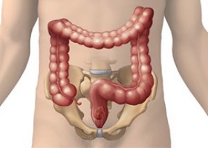
Rectal diseases are characterized by typical symptoms that develop during the development of pathological processes in it and the anal canal. In the practice of a surgeon, rectal diseases are common, but the disease can be caused not only by rectal pathology, which is dealt with by surgeons and proctologists, but also by oncological processes, urological and gynecological pathology.
Diseases of the rectum manifest themselves polymorphically, but the main symptoms are quite clear: pain in the anus, often radiating to the sacrum and coccyx, perineum, thigh, to one degree or another, a violation of the act of defecation and pain during it; itching; there may be discharge, bleeding.
 [
[ How to recognize rectal diseases?
There are quite a lot of diagnostic methods, a proctologist conducts a more detailed diagnosis, but a surgeon should refer a fully examined patient to a specialist. In most cases, there is no need to refer to a proctologist, except for indications for surgical treatment. The main diagnostic method that will allow identifying diseases of the rectum is rectal: examination, digital examination, anoscolia using a rectal mirror, rectoscopy, fibrocolonoscopy. It is necessary to conduct a stool analysis: coproscopy, stool analysis for helminth eggs, occult blood, dysbacteriosis, in the presence of discharge - cytology and microflora analysis. If you doubt the primacy of the pathology or its connection with other pelvic organs, a consultation with a gynecologist and urologist is necessary.
Rectal examination allows to identify the functional state of the muscles of the anus, pathological changes in the tissues of the anal canal (cracks, fistulas, hemorrhoids, hypertrophy of the anal papillae, fibrous polyps, benign and malignant tumors, post-traumatic cicatricial changes, foreign bodies, intestinal stenosis), cystic and tumor-like formations, inflammatory infiltrates in the pararectal tissue, changes in the prostate gland and seminal vesicles in men (adenoma, cancer, prostatitis) and genitals in women, peritoneum, Douglas space.
The diagnostic procedure begins with an examination of the anus: skin, position of the anus (its displacement indicates the presence of a rectal disease in the pararectal tissue: paraproctitis, abscess, tumor, cicatricial displacement in chronic proctitis), the locking mechanism (at rest and during straining), the presence of visible changes - malformations (prolapse, stenosis), traumatic injuries, cracks, hemorrhoids, fistulas, polyps, cicatricial changes, hypertrophy of the papillae, etc.
Digital examination is carried out gradually with the patient lying on his side, on his back, or better yet, in the knee-elbow position. First, the tone, extensibility, elasticity of the external anal sphincter, mucous membrane, presence and degree of pain during the procedure are examined. Then the finger is advanced into the ampulla itself, determining the state of its lumen (expansion, narrowing, presence of foreign bodies), carefully palpating and assessing the walls (elastic and soft in the norm; pasty, presence of bulges, seals, cicatricial deformations), the state of the prostate in men, the rectovaginal wall and the cervix in women are determined; the state of the pararectal tissue, the adjacent wall of the Douglas space, the inner surface of the sacrum and coccyx are determined. After removing the finger, the glove is examined for the presence of discharge: normal feces, altered feces, mucous, bloody, purulent, ichorous discharge. To study the upper rectal region and pelvirectal tissue, a bimanual examination is used.
Anoscopy with a rectal mirror helps to identify rectal diseases and anal pathology. Rectomanoscopy and colonoscopy provide in-depth results (detection of cancer, polyposis, non-specific ulcerative colitis, examination of deep fistulas and other pathologies). For this, the patient must be sent prepared (castor oil 30 g the day before; enemas in the evening and in the morning until clear water) to an endoscopist.