
All iLive content is medically reviewed or fact checked to ensure as much factual accuracy as possible.
We have strict sourcing guidelines and only link to reputable media sites, academic research institutions and, whenever possible, medically peer reviewed studies. Note that the numbers in parentheses ([1], [2], etc.) are clickable links to these studies.
If you feel that any of our content is inaccurate, out-of-date, or otherwise questionable, please select it and press Ctrl + Enter.
Wedge-shaped defect of dental hard tissues: what to do, how to treat, restoration
Medical expert of the article

Last reviewed: 04.07.2025
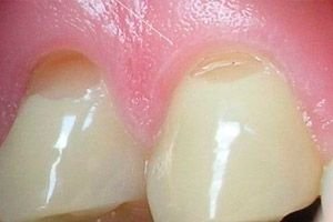
A specific form of dental pathology – a wedge-shaped tooth defect – is related to non-carious enamel damage. This defect occurs at the neck of the tooth in its visible area. The upper part of the “wedge” in all cases “looks” into the dental cavity.
This type of damage is found mainly in patients after 30-45 years of age and is located symmetrically on the teeth of only the upper or only the lower jaw.
Epidemiology
Statistical information concerning such a pathology as a wedge-shaped defect varies greatly. This can be explained by some inconsistencies in this term. Thus, specialists who consider any cervical damage to enamel as a type of wedge-shaped defect indicate that the disease occurs in almost 85% of patients in dental clinics. However, such a figure can hardly correspond to reality.
Another category of dentists bases their statistics solely on the registration of clear and deep cervical lesions. According to their data, the disease is detected in only 5% of patients.
One can only guess which information is closer to the truth.
It has been noted that the disease affects mainly men. Right-handed people often have a problem on the right side of the dental arch, while left-handed people often have a problem on the left side.
Among all the teeth, premolars are the ones that suffer most from the disease.
Causes wedge-shaped dental defect
The exact causes of the disease have not been determined to date. Experts have identified individual risk factors that can trigger the development of the pathology. We are talking about the following factors:
- Violation of the integrity of the enamel when using rough and hard dental accessories, as well as when cleaning teeth incorrectly. The point is that near the neck, the enamel coating is especially thin, so with strong mechanical friction it wears out faster.
- Demineralization processes. The accumulation of plaque in the cervical area leads to the fact that bacteria producing acid begin to actively multiply in it. The acid, in turn, destroys the calcium present in the enamel coating of the tooth.
- Increased load on the cervical area of individual teeth. This factor is associated with malocclusion and incorrect jaw movements when chewing food.
- Wearing braces.
Less often, the "culprits" are diseases that are accompanied by frequent heartburn and vomiting. The mechanism of disease development in such situations is clear: acid from the stomach, getting into the oral cavity, accumulates near the gum and gradually "corrodes" dental tissue.
 [ 6 ]
[ 6 ]
Pathogenesis
The pathogenetic characteristic of the disease is the gradual damage of the enamel coating. The damage does not occur immediately and goes through several stages:
- The initial stage, when changes in the enamel are not "obvious" during a normal examination of the oral cavity. Sometimes the patient may note the presence of tooth sensitivity or slight clouding of the enamel.
- The middle stage is accompanied by pronounced sensitivity of the affected teeth (for example, to high and/or low temperatures, acidic foods, etc.). At this stage, slow destruction of tissues begins.
- Progress stage: for this stage, the appearance of a deep defect is typical – from 2 to 4 mm. A characteristic “wedge” with a pointed top becomes noticeable.
- Deep stage: the depth of the defect exceeds 4 mm. Dentin may be affected.
Symptoms wedge-shaped dental defect
The main difficulty for dentists is timely recognition of the disease. The fact is that a person does not immediately feel the presence of pathology: there is no pain, the affected area is covered by the gum and is not visible.
The first signs may appear only when the disease has progressed to the third or even fourth stage.
Dentists advise paying attention to the following symptoms in a timely manner:
- tooth pigmentation, clouding and paleness of enamel;
- exposure of the tooth neck, change in the boundaries of the gum in relation to the tooth;
- discomfort and hypersensitivity of individual teeth.
A wedge-shaped defect of tooth enamel can affect one tooth or several, usually located in one row. The wedge-shaped cavity does not turn black, as with caries: its walls are smooth and hard. The dental cavity in all cases remains closed (that is why the patient does not feel any pain).
A wedge-shaped defect of the hard tissues of the tooth always develops only in the cervical zone and on the anterior surface of the enamel.
The development of the disease can begin with almost any tooth, both maxillary and mandibular. Most often, premolars, canines and first molars are affected - mainly due to their protruding position. A wedge-shaped defect of the front teeth is also possible, but somewhat less often.
Wedge-shaped defect of teeth in children is observed extremely rarely: to date, only isolated cases of such pathology in pediatric patients are known.
Complications and consequences
Damage to dentin in the cervical area can lead to the following complications:
- to the inflammatory process in the pulp;
- to dystrophic changes in the pulp;
- to periodontitis;
- to increased sensitivity of gums and teeth.
In cases where dentin is deeply damaged, a pathological fracture of the tooth crown may occur.
With a long-term "wedge" recessive processes in the gums may occur. This, in turn, can cause loosening of the teeth, as well as damage to the periodontium.
The main consequence that worries most patients with such a defect is the unacceptable aesthetic appearance of the teeth.
Diagnostics wedge-shaped dental defect
The disease can usually be easily identified by visual examination. However, before starting treatment, the doctor may prescribe certain types of examinations and tests. For example, an X-ray examination is often prescribed.
During a visual examination of the oral cavity, the doctor discovers a tooth defect in the form of a wedge (V-shaped cut, or step). The defect has smooth borders, a dense bottom and glossy walls.
It is not necessary to determine the composition of the gingival fluid in case of a wedge-shaped tooth defect, but some patients still undergo this type of analysis. Gingival fluid is a physiological mass that fills the gingival groove. Several methods are used to obtain this fluid:
- gingival wash;
- using a micropipette;
- insertion of a special absorbent paper strip into the groove.
The composition of the fluid is usually represented by bacteria and their waste products, elements of blood serum, intercellular fluid of gum tissue, and leukocytes.
The composition may change with the development of periodontal diseases and inflammatory processes.
Tests are rarely prescribed in dental practice. In some cases, if there is an inflammatory process of unclear etiology, the patient is offered to take a general blood test, as well as a discharge test (if any).
Instrumental diagnostics in the vast majority of cases consists of conducting an X-ray examination. The essence of the method is to obtain a local X-ray image of the affected areas using a radiovisiograph. The image is obtained thanks to X-rays. Targeted radiography allows you to pay attention to many dental features: using this method, you can diagnose hidden caries, periodontal pathologies, and examine the condition of the dental canals.
Computer tomography is used relatively rarely, only when it is necessary to obtain a three-dimensional image. The method allows for a thorough assessment of the condition of the teeth, periodontium, sinuses, temporomandibular joint, etc.
The procedure of electroodontodiagnostics is carried out when it is necessary to assess the viability of the dental pulp. This method will help to determine which tooth tissues are affected by the painful destructive process, as well as to assess the need for intervention in the root canals.
Differential diagnosis
The overwhelming majority of cases with a wedge-shaped defect do not require differential diagnostics, as they have characteristic distinguishing features. Differentiation is carried out only in certain situations.
- Wedge-shaped defect and caries.
The "wedge" is always localized in the cervical part of the tooth and has a typical shape corresponding to the name of the disease, and also has a hard and smooth wall. The carious cavity is filled with soft darkened dentin, which is accompanied by unpleasant sensations from the effects of irritants.
- Wedge-shaped defect and erosions.
The erosion is cup-shaped and is located on the entire anterior surface of the tooth. Increased sensitivity and darkening of the dentin are usually absent.
- Wedge-shaped defect and post-acid necrosis.
Post-acid necrosis is localized on the front teeth: the enamel coating becomes uneven and grayish-dirty, loses its smoothness and shine. The teeth become sensitive and brittle, with their gradual destruction.
Who to contact?
Treatment wedge-shaped dental defect
Regardless of the stage of development of the defect, the doctor will first prescribe treatment aimed at eliminating the provoking factor: they treat the digestive system, correct malocclusion, etc.
Next, they begin to eliminate the defect itself. At the initial stage of the pathology development, application of preparations that give calcium and fluoride to dental tissues can help. Such procedures are called calcination and fluoridation. It is advisable to carry them out in courses, twice a year: this stops destructive processes and restores surface enamel.
At home, you can use special varnish and gel coatings, which are applied according to the scheme specified by the doctor. It is recommended to brush your teeth with special pastes - this must be done regularly, for a long time.
At other stages of defect development, procedures will be required to correct the aesthetic appearance of the affected teeth.
Tooth restoration with a wedge-shaped defect
The filling is installed using filling materials that are highly elastic. The area near the neck is always subject to heavy loads, so a regular filling will inevitably fall out sooner or later. To ensure that the filling holds well, special notches are made on the surface of the defect.
The filling is a fluid mass with a high degree of elasticity, which is applied using a syringe and polymerized with a special lamp.
Additional protection of the neck and improvement of the aesthetic appearance of the affected teeth can be achieved with veneers or microprostheses. Veneers are thin ceramic plates that cover the dental defect. The disadvantages of such restoration include the importance of periodic replacement of microprostheses. Although, today, there are veneers that can last up to two decades.
Another method of restoration is dental crowns. They, like veneers, do not prevent further destruction of layers. For this, it is necessary to carry out appropriate treatment aimed at eliminating the original cause of the defect.
How to close a wedge-shaped defect on a lateral tooth or on other damaged teeth? Considering the above, we can highlight the following main options:
- filling;
- installation of microprostheses;
- installation of crowns.
Is it necessary to treat wedge-shaped defect of teeth?
Treatment of the defect is necessary. And not only to eliminate unpleasant symptoms, but also to block further aggravation of the disease.
- Fluoridation of teeth is the application of fluoride-containing preparations to the affected areas of the teeth, which promotes tissue restoration. Additionally, increased sensitivity is eliminated.
- Calcification is the treatment of damaged enamel with calcium preparations, which stops the further development of the disease.
- Laser treatment is the treatment of the defect with a laser. This procedure provides enamel compaction and eliminates increased sensitivity.
If treatment is not carried out, then dental prosthetics or installation of crowns will only provide a temporary solution to the problem. In the future, the disease will worsen, which can lead to the fracture of the affected tooth in the area of damage.
[ 20 ], [ 21 ], [ 22 ], [ 23 ]
Treatment at home
In addition to the necessary dental treatment, you can also try folk remedies. For example, there are a number of methods that should improve the condition of patients with a wedge-shaped defect:
- Buy an alcohol tincture of propolis at the pharmacy, dilute a few drops in a glass of warm water. Use this water for rinsing after meals.
- They try to regularly include kelp, parsley, basil, and iodized salt (in the absence of contraindications) in their diet.
- Sea mother-of-pearl shells are ground to a powder. The resulting powder is applied to the teeth with a brush and held there for as long as possible without rinsing the mouth.
- Apply lemon or lime leaves to the affected teeth.
- Include grated horseradish in your diet.
- Lubricate the teeth and gums with a mixture of honey and cinnamon powder.
In addition, it is useful to regularly include foods with sufficient mineral content in your meals. For example, calcium can be obtained from dairy products, and fluorine from seaweed, beans, chicken, buckwheat, bananas, citrus fruits, and honey.
Toothpaste for wedge-shaped defect of teeth
Dentists advise choosing toothpastes with a desensitizing effect for brushing teeth:
- ROCS Medical minerals (remineralizing paste), there is a version for adult patients and children. The product reduces the sensitivity of tooth tissues.
- ROCS Medical sensitive will help eliminate discomfort and pain.
- Doctor Best Sensitive or Elmex Sensitive are fluoride-containing, with reduced abrasive properties.
There are also a number of toothpastes that help with wedge-shaped defects:
- Bio Repair;
- Sensigel;
- Oral-B sensitive fluoride;
- Biodent sensitive.
To achieve the effect, any of the listed pastes should be used regularly. Only the attending dentist can accurately determine the duration of use of such products.
Irrigator for wedge-shaped defect and sensitive teeth
An irrigator is a device that facilitates oral care. It delivers a stream of water or medicine, washing the teeth, the space between the teeth, which serves as a good prevention of caries, periodontal disease and plaque formation. Simultaneous gum massage improves local blood circulation.
The use of an irrigator is especially recommended:
- with frequent inflammatory processes in the oral cavity, with bleeding gums;
- when wearing braces;
- if you have bad breath;
- in diabetes mellitus.
An irrigator can serve as a preventative measure against wedge-shaped defects. If the disease already exists, then this device can be used to prevent further development of the disease. Contrary to the opinion of many, an irrigator does not aggravate the problem of dental defects, but it is not able to cure them either.
Why do teeth hurt after treatment of wedge-shaped defects?
Toothache after treatment is not a typical situation. It happens relatively rarely and can be associated with several factors:
- the presence of additional dental problems (caries, damage to dentin and pulp);
- hypothermia, upper respiratory tract diseases;
- poor quality filling, development of inflammation at the site of filling installation.
The pain may persist throughout the day, becoming more severe at night.
Often, pain is associated with individual hypersensitivity of the patient, with increased tone of the vagus nerve, with high blood pressure, with irritation of the trigeminal nerve, as well as with otolaryngological pathologies (for example, inflammation of the nasal sinuses).
Normally, teeth should not hurt after treatment. If pain is present, then diagnostics should be performed to determine the source of the pain.
Prevention
In order to prevent the occurrence of pathology, it is very important to monitor your own health in general, promptly seek medical help when necessary. This applies to both dental problems and malfunctions in the work of other organs and systems.
In addition, it is equally important to adhere to the basic rules of oral hygiene:
- teeth should be brushed in the morning after breakfast and at night after the last meal;
- It is advisable to choose a toothbrush with medium-hard bristles;
- It is important to remember that after each meal you should rinse your mouth;
- It is necessary to eliminate any excessive mechanical stress on the teeth: do not crack nut shells, chew threads, etc.
A timely consultation with a dentist will help to detect the disease at an early stage of formation. This will allow eliminating the pathology with simpler and more accessible means, which will be less painful and less costly financially.
Forecast
Wedge-shaped dental defect is considered a relatively safe dental pathology. However, this does not mean that the patient can ignore it. The disease needs to be treated, and the sooner, the better for the patient. If the pathology is neglected, the treatment will be more difficult and radical.