
All iLive content is medically reviewed or fact checked to ensure as much factual accuracy as possible.
We have strict sourcing guidelines and only link to reputable media sites, academic research institutions and, whenever possible, medically peer reviewed studies. Note that the numbers in parentheses ([1], [2], etc.) are clickable links to these studies.
If you feel that any of our content is inaccurate, out-of-date, or otherwise questionable, please select it and press Ctrl + Enter.
Crouzon syndrome
Medical expert of the article

Last reviewed: 04.07.2025
 [
[ Causes Crouzon syndrome
To date, a fairly large number of various clinical and laboratory studies of the causes of this disease have been conducted. Scientists have come to the unambiguous conclusion that the syndrome has an autosomal dominant path of genetic inheritance.
This means that if the mutation gene is present in one parental chain (maternal or paternal), there is a 50% risk of having a baby with signs of Crouzon syndrome.
Children do not always inherit a damaged chromosome. Moreover, they may not even be carriers of the defect. Therefore, parents, one of whom has a mutated gene in the family, have every chance of having a healthy child. The main thing is to undergo a thorough examination before planning a pregnancy.
Thus, the following risk factors can be identified:
- the presence of Crouzon syndrome in one of the parents or in blood relatives;
- carriage of a mutated gene by one of the parents;
- the father's age is over 60 years (at the time of conception of the child).
Pathogenesis
The pathogenesis of the syndrome is simple: the disorder is provoked by a gene mutation of the fibroblast growth factor FGFR2. This gene is located in a specific chromosome (10q26) and consists of 20 sections with gene information. The change that leads to the appearance of Crouzon syndrome is most often located in sections of the seventh and ninth gene.
In total, the FGFR2 gene can contain 35 mutational changes that affect the development of the syndrome. Most often, this disorder occurs on the paternal side.
All small children have sutures - small spaces between the elements of the bones of the cranium and face. As the baby grows and develops, its brain increases, and thanks to these sutures, the corresponding expansion of the skull occurs. The suture spaces grow together only when the brain is fully formed and stops growing.
In children with Crouzon syndrome, the sutures close much earlier than necessary. Therefore, the growing brain is forced to "adapt" to the space that is available. Externally, this becomes noticeable by the non-standard shape of the skull, face and teeth.
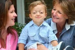
Symptoms Crouzon syndrome
The first signs of the syndrome are visible immediately after the birth of the child. They can be observed in the area of the face and cranium:
- change in the shape of the middle part of the face;
- change in the shape of the nose;
- protruding tongue;
- shortened and low-set lip;
- insufficient jaw closure.
- The skeletal system undergoes changes. The following types of cranial deformation may appear:
- trigonocephaly - a wedge-shaped head with an expanded occipital and narrowed frontal part;
- scaphocephaly - a boat-shaped head with an elongated, low-set skull and a narrow forehead;
- brachycephaly - short head, or too wide head with a shortened skull length;
- Kleeblattschadel defect is a hydrocephaloid deformity of the skull in the shape of a trefoil.
By palpation, you can feel the flat sutures on the skull. But this is not always possible, since the sutures can heal at any stage:
- at the stage of embryonic development;
- in the first year of the baby's life;
- closer to 3 years;
- up to 10 years of age.
- There are disturbances in the visual organs:
- primary or secondary exophthalmos - protrusion of the eyes, in which the eyeball remains unchanged;
- nystagmus – frequent involuntary vibrations of the eyeballs;
- multilateral strabismus - incorrect divergent position of the eyes;
- hypertelorism - an enlarged space between the inner corners of the eyes and the pupils;
- ectopia – deviation of the pupil or lens from the center;
- coloboma – absence of part of the iris;
- megalocornea - pathological enlargement of the cornea.
- Defects of the auditory organs are also noted:
- conductive deafness;
- change in the shape of the internal auditory canal;
- decreased sound conductivity of bones;
- atresia of the external auditory canal.
As can be seen from the clinical picture, all symptoms of the disease are localized only in the head area. It is characteristic that no vestibular disorders are observed.
Complications and consequences
Crouzon syndrome cannot pass without a trace: as a rule, the child is left with various consequences and complications:
- hydrocephalus;
- deterioration of vision, even loss (as a result of prolonged compression of the optic nerve, irreversible changes occur in it);
- thinning and ulcerative damage to the cornea (due to excessive bulging of the eyeballs, it becomes impossible to completely close the eyelids, as a result of which the cornea partially dries out and becomes covered with ulcers);
- mental retardation;
- difficulties with adaptation in society (mental disability and unpleasant external manifestations of the syndrome significantly complicate the patient’s interaction with society).
Another complication of the syndrome may be Arnold-Chiari malformation, which is the displacement of the cerebellar tonsils through the foramen magnum to the cervical vertebrae.
Diagnostics Crouzon syndrome
First of all, the doctor examines the sick child. He can clarify whether something similar has happened in the family, because the signs of Crouzon syndrome are quite characteristic, and it is difficult to confuse them.
Instrumental diagnostics, which are mandatory and carried out immediately upon suspicion of the syndrome, will help the doctor to clarify the diagnosis.
Radiography will indicate the stage of fusion of the lambdoid, coronal and sagittal sutures. In addition, this method helps to detect a decrease in the paranasal sinuses, signs of basilar kyphosis, an enlarged pituitary fossa, and an irregular shape of the orbits.
Topographically, deformation of the internal auditory canal is observed. Also, in the topogram of the temples, one can trace the external rotation of the petrous part of the pyramid, which occurs against the background of dysplasia of the base of the skull. Visually, this is manifested by hyperostosis, oblique direction of the auditory canals, and incorrect course of the facial nerve.
Computed tomography or MRI confirms the following signs:
- atresia;
- narrowing of the external auditory canal;
- deformation of the chambers of the mastoid process and stapes;
- absence of the tympanic cavity;
- ankylosis of the malleus;
- disruption of the development of the periosteal region of the labyrinth.
Additionally, the doctor may refer the patient to other specialists who will prescribe tests and other studies at their discretion. For example, if Crouzon syndrome is suspected, consultations with a geneticist, psychiatrist, neurologist, ophthalmologist, and neurosurgeon are appropriate.
Differential diagnosis
Differential diagnosis includes isolated craniosynostosis, Apert syndrome, Saethre-Chotzen syndrome and Pfeiffer syndrome.
Who to contact?
Treatment Crouzon syndrome
Unfortunately, it is impossible to completely cure Crouzon syndrome. Treatment can be aimed at functional and cosmetic correction: this can only be achieved surgically. During the operation, the surgeon partially opens the synostotic sutures and also corrects the position of the eyeball.
Let us describe in more detail the process of surgical treatment of Crouzon syndrome. Such treatment is best performed at the age of 4-5 years. Thanks to the operation, maxillary hypoplasia is corrected, the dentition is restored and exophthalmos is removed (the lower edge of the eye sockets moves forward and increases in volume). During the intervention to establish the bite, the doctor fixes the jaws with special plates, which will be removed only after 1-1.5 months.
Modern medicine uses distraction methods to correct facial bone deformations. There are special devices for shifting almost any part of the skull, both from the front and from the back of the head. Such treatment is only just gaining momentum, and over time, we can hope that the correction of bone defects will become more gentle and effective.
Drug therapy is not the main treatment for Crouzon syndrome. Thus, drugs can be used only to alleviate the patient's condition.
Nootropic drugs |
||
Piracetam |
Pantogam |
|
Method of administration and dosage |
Usually 30-50 mg of Piracetam per day is prescribed. The treatment is long-term. |
The daily dose of the drug for Crouzon syndrome can range from 0.75 to 3 g. The duration of therapy is up to 4 months (sometimes longer, at the discretion of the doctor). |
Contraindications |
Renal failure, diabetes mellitus, children under 1 year of age. |
Acute renal failure, phenylketonuria, tendency to allergies. |
Side effects |
Overexcitement, irritability, sleep and appetite disorders, headaches. |
Allergies, sleep disorders, tinnitus. |
Special instructions |
It is not recommended to take more than 5 g of the drug per day. |
If the treatment is long-term, then Pantogam is not recommended to be combined with other nootropic drugs. |
Vascular drugs |
||
Cavinton |
Cinnarizine |
|
Method of administration and dosage |
For Crouzon syndrome, a long course of treatment is practiced with the intake of 5-10 g of the drug three times a day. |
The drug is taken for a long time, 75 mg per day. |
Contraindications |
Severe heart disease, heart rhythm disturbances, unstable blood pressure. |
Allergic tendency. |
Side effects |
Increased heart rate, decreased blood pressure. |
Sleep disorders, dyspepsia. |
Special instructions |
The drug cannot be combined with heparin. |
The drug enhances the effect of sedatives. |
Diuretics |
||
Lasix |
Diacarb |
|
Method of administration and dosage |
The treatment regimen for Crouzon syndrome is individual and depends on the indications. |
Prescribed on average 0.25 g 1-4 times a day. |
Contraindications |
Kidney dysfunction, hypokalemia, dehydration, difficulty urinating, tendency to allergies. |
Acidosis, diabetes mellitus. |
Side effects |
Muscle weakness, cramps, headache, arrhythmia, hypotension. |
Drowsiness, fatigue, headache, anemia. |
Special instructions |
During treatment, constant medical supervision is necessary. |
Should not be used for long periods of time. |
Prevention
Prevention of the birth of children with Crouzon syndrome is impossible, due to the fact that this disease is hereditary in the vast majority of cases.
Since sporadic cases of the syndrome do sometimes occur, which may be associated with the advanced age of the child's father at the time of conception, it is recommended to carefully weigh the degree of risk when planning such a "late" pregnancy.
If there have already been cases of children born with Crouzon syndrome in the family, then it makes sense for parents to undergo a full examination by a geneticist for the presence of a mutated FGFR2 gene.
All pregnant women, regardless of the quality of their heredity, are recommended to register with a women's clinic in a timely manner (no later than 12 weeks), and also regularly visit a gynecologist.
Forecast
Unfortunately, even after successful surgery, no one can 100% guarantee positive dynamics of such a disease as Crouzon syndrome. Often, the patient's visual function is completely or partially impaired due to atrophic changes in the optic nerve. Due to the irregular shape of the eye sockets, problems with holding the eyeball arise. Over time, bone defects become more pronounced.
However, many patients still manage to be socially adapted for a long time, regardless of the degree of manifestation of the disease. It remains to be hoped that the level of medicine is progressing, and in the near future there will still be methods that allow us to prevent and treat any gene disorders, including Crouzon syndrome.