
All iLive content is medically reviewed or fact checked to ensure as much factual accuracy as possible.
We have strict sourcing guidelines and only link to reputable media sites, academic research institutions and, whenever possible, medically peer reviewed studies. Note that the numbers in parentheses ([1], [2], etc.) are clickable links to these studies.
If you feel that any of our content is inaccurate, out-of-date, or otherwise questionable, please select it and press Ctrl + Enter.
Caseous pneumonia
Medical expert of the article

Last reviewed: 07.07.2025
Caseous pneumonia is one of the most severe forms of pulmonary tuberculosis. It is characterized by a pronounced caseous-necrotic component of tuberculous inflammation, rapid progression and the formation of multiple cavities of decay. It can occur as an independent disease in a previously healthy person or as a complication of another form of pulmonary tuberculosis. There are two clinical forms of caseous pneumonia: lobar and lobular. Lobar caseous pneumonia usually develops as an independent clinical and anatomical form of tuberculosis, and lobular often complicates other forms of pulmonary tuberculosis.
 [
[ Epidemiology of caseous pneumonia
Against the background of social and economic upheavals, frequent disorganization in the work of the anti-tuberculosis service, the number of patients with this form of tuberculosis has increased. Caseous pneumonia was again included in the Russian clinical classification of tuberculosis. In recent years, caseous pneumonia has been observed in 3-5% of newly diagnosed patients with tuberculosis. Adults from medical and social risk groups associated with the risk of developing immunodeficiency (HIV-infected, alcoholics, socially maladjusted individuals, as well as those treated for a long time with glucocorticoids, cytostatic drugs, etc.) are most susceptible to caseous pneumonia. An important factor increasing the risk of developing caseous pneumonia is considered to be human infection with highly virulent, drug-resistant Mycobacterium tuberculosis.
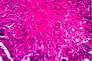
Pathogenesis and pathological anatomy of caseous pneumonia
The occurrence of caseous pneumonia is associated with intensive reproduction of mycobacteria in the lung tissue, which occurs against the background of severe immunodeficiency. metabolic failure of phagocytic cells and lymphocytes. Pathological increase in apoptosis of cells involved in the immune response is considered the main pathogenetic factor in the development of caseous pneumonia.
The initial stage of caseous pneumonia (acinous, acinous-lobular, confluent lobular) is characterized by massive cell death in the affected area and the formation of an extensive zone of caseous necrosis. The pathological process quickly moves to the next, more widespread and irreversible stage. Caseous foci and foci are formed in the adjacent lung tissue, merging with each other. Mycobacteria penetrate into the lumen of small bronchi, lymphatic and blood vessels. Their spread and progression of caseous changes over 2-3 weeks lead to widespread lung damage. A morphological feature of caseous pneumonia is considered to be a sharp predominance of caseous-necrotic changes over other specific changes in the lung tissue.
In the mechanism of lung tissue decay, the damaging effect of the pathogen's waste products is of great importance, causing macrophage cytolysis and the entry of lysosomal enzymes, prostaglandins and TNF-α into the lung tissue. Significant microcirculation disorders caused by necrotic vasculitis also contribute to the decay of lung tissue. Melting of caseous masses leads to the formation of multiple cavities of various sizes - acute caverns. The destructive process in the lung is accompanied by a temporary increase in the partial oxygen pressure in the affected area, which creates optimal conditions for the intensive reproduction of mycobacteria.
Without treatment, caseous pneumonia often leads to death. The cause of death is pulmonary heart failure, developing against the background of destruction of lung tissue and severe intoxication.
With timely initiation of complex treatment, the rapid progression of the process can be stopped. The gradual organization of fibrinous masses causes the appearance of carnification areas: cavities are transformed into fibrous caverns, caseous-necrotic foci are encapsulated. Thus, caseous pneumonia, in which changes in the lungs are largely irreversible, is transformed into fibrous-cavernous tuberculosis of the lungs.
Symptoms of caseous pneumonia
Typical caseous pneumonia develops acutely. In the initial stage, when caseous-necrotic masses form in the affected area, intoxication syndrome is expressed (fever, chills, weakness, severe sweating, sharp deterioration of appetite), shortness of breath, cough, mostly dry, sometimes with a small amount of difficult to separate sputum.
After the melting of caseous-necrotic masses and the formation of multiple cavities of decay in the lung, the severity of the bronchopulmonary-pleural syndrome increases sharply. The cough becomes wet, with a large amount of sputum. Patients are bothered by chest pain. Blood may appear in the sputum. Dyspnea increases, acrocyanosis develops. Hectic fever of the wrong type is noted, often the development of cachexia.
During physical examination, shortened percussion sounds are detected over the affected areas of the lung, weakened bronchial breathing and moist fine-bubble rales are heard. After the formation of cavities of decay, the rales become sonorous, numerous, medium- and large-bubble. The appearance of tachycardia and accentuation of tone II over the pulmonary artery are noted. An enlarged liver is often observed.
Where does it hurt?
What's bothering you?
X-ray picture of caseous pneumonia
X-ray examination of the chest organs reveals widespread gross changes. In patients with lobar caseous pneumonia, darkening of the entire or greater part of the lung lobe is determined, initially homogeneous. As the disease progresses, areas of enlightenment of an irregular bay-like shape with unclear contours appear. On CT ("air bronchography"), the lumens of the dilated medium and large bronchi can be clearly distinguished in the compacted lobe of the lung. Later, as the caseous masses are rejected, the cavities acquire the characteristic features of a cavern with a gradually forming wall. In the adjacent segments and in the other lung, foci of bronchogenic seeding are often visible. The affected lobe of the lung decreases as a result of loss of elasticity.
In lobular caseous pneumonia, large focal shadows and small foci with a diameter of about 1.5 cm are visible on the X-ray in direct projection. The shadows have an irregular shape, medium or high intensity, and unclear contours. Tomography reveals multiple decay cavities in the lungs).
What do need to examine?
How to examine?
Who to contact?
Treatment of caseous pneumonia
Treatment of caseous pneumonia is carried out using anti-tuberculosis drugs.