
All iLive content is medically reviewed or fact checked to ensure as much factual accuracy as possible.
We have strict sourcing guidelines and only link to reputable media sites, academic research institutions and, whenever possible, medically peer reviewed studies. Note that the numbers in parentheses ([1], [2], etc.) are clickable links to these studies.
If you feel that any of our content is inaccurate, out-of-date, or otherwise questionable, please select it and press Ctrl + Enter.
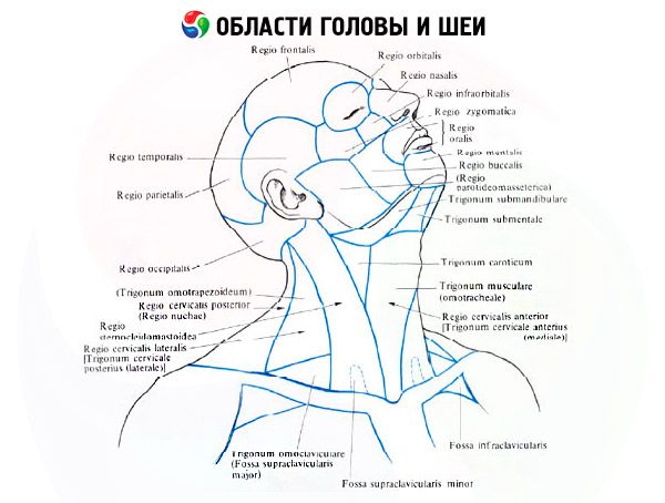
Areas and triangles of the neck
Medical expert of the article

Last reviewed: 06.07.2025
The upper border of the neck is drawn (on the right and left) from the chin along the base and posterior edge of the branch of the lower jaw to the temporomandibular joint, continuing backwards through the apex of the mastoid process of the temporal bone along the superior nuchal line to the external protrusion of the occipital bone.
The lower border of the neck runs on each side from the jugular notch of the sternum along the upper edge of the clavicle to the apex of the acromion and further to the spinous process of the seventh cervical vertebra.
Taking into account the relief of the skin on the neck, determined by the position of the deeper muscles and internal organs, the following areas of the neck are distinguished in the anterior sections: anterior, sternocleidomastoid (right and left) and lateral (right and left), as well as posterior.
The anterior region of the neck, or the anterior triangle of the neck (regio cervicalis anterior, s.trigonum cervicale anterius), is bounded on the sides by the sternocleidomastoid muscles. At the top, the base of the triangle is formed by the lower jaw, and its apex reaches the jugular notch of the manubrium of the sternum.
In the anterior region of the neck, in turn, a medial triangle of the neck is distinguished on each side, limited in front by the median line, above by the lower jaw and behind by the anterior edge of the sternocleidomastoid muscle. A conditional horizontal plane drawn through the body and the greater horns of the hyoid bone divides the median region of the neck (anterior triangle) into two regions: the upper suprahyoid (regio suprahyoidea) and the lower sublingual (regio unfrahyoidea). In the sublingual region of the neck, two triangles are distinguished on each side: the carotid and muscular (scapular-tracheal).
The carotid triangle (trigonum caroticum) is bounded above by the posterior belly of the digastric muscle, behind by the anterior edge of the sternocleidomastoid muscle, and in front and below by the superior belly of the omohyoid muscle. Within this triangle, above the superficial plate of the cervical fascia, are the cervical branch of the facial nerve, the superior branch of the transverse nerve of the neck, and the anterior jugular vein. Deeper, under the superficial plate of the cervical fascia, are the common carotid artery, the internal jugular vein, and behind them, the vagus nerve, enclosed in a sheath of the vascular-nerve bundle common to them. The deep lateral cervical lymph nodes also lie here. Within the carotid triangle, at the level of the hyoid bone, the common carotid artery divides into the internal and external carotid arteries. The branches that branch off from the latter are the superior thyroid, lingual, facial, occipital, posterior auricular, ascending pharyngeal arteries and the sternocleidomastoid branches, which go to the corresponding organs. Here, in front of the sheath of the vascular-nerve bundle, is the superior root of the hypoglossal nerve, deeper and lower is the laryngeal nerve (a branch of the vagus nerve), and even deeper on the prevertebral plate of the cervical fascia is the sympathetic trunk.
The muscular (scapulotracheal) triangle (trigonum musculare, s. omotracheale) is limited posteriorly and inferiorly by the anterior edge of the sternocleidomastoid muscle, superiorly and laterally by the superior belly of the omohyoid muscle, and medially by the anterior midline. Within this triangle, immediately above the jugular notch of the manubrium of the sternum, the trachea is covered only by the skin and the fused superficial and pretracheal plates of the cervical fascia. Approximately 1 cm to the side of the midline is the anterior jugular vein, which goes into the suprasternal interfascial cellular space.
In the suprahyoid region, three triangles are distinguished: the submental (unpaired) and the paired ones - the submandibular and lingual.
The submental triangle (trigonum submentale) is bounded on the sides by the anterior bellies of the digastric muscles, and its base is the hyoid bone. The apex of the triangle faces upward, toward the mental spine. The bottom of the triangle is the right and left mylohyoid muscles, connected by a suture. The submental lymph nodes are located in the area of this triangle.
The submandibular triangle (trigonum submandibulare) is formed at the top by the body of the lower jaw, and at the bottom by the anterior and posterior bellies of the digastric muscle. The submandibular salivary gland is located here. The cervical branch of the facial nerve and the branching of the transverse nerve of the neck penetrate this triangle. The facial artery and vein are also located superficially here, and the retromandibular vein is located behind the submandibular gland. Within the submandibular triangle, the lymph nodes of the same name are located under the lower jaw.
The lingual triangle (Pirogov's triangle) is small but very important for surgery, located within the submandibular triangle. Within the lingual triangle is the lingual artery, access to which is possible in this place of the neck. In front, the lingual triangle is limited by the posterior edge of the mylohyoid muscle, behind and below - by the posterior belly of the digastric muscle, and above - by the hypoglossal nerve.
In the lateral region of the neck, the scapuloclavicular and scapulotrapezoid triangles are distinguished.
The scapuloclavicular triangle (trigonum omoclaviculare) is located above the middle third of the clavicle. It is limited below by the clavicle, above by the lower belly of the omohyoid muscle, and in front by the posterior edge of the sternocleidomastoid muscle. In the area of this triangle, the terminal (third) part of the subclavian artery, the subclavian part of the brachial plexus, between the trunks of which the transverse artery of the neck passes, and above the plexus - the suprascapular and superficial cervical arteries. Anterior to the subclavian artery, in front of the anterior scalene muscle (in the prescalene space), lies the subclavian vein, firmly fused with the fascia of the subclavian muscle and the plates of the cervical fascia.
The scapulotrapezoid triangle (trigonum omotrapezoideum) is formed by the anterior edge of the trapezius muscle, the lower belly of the omohyoid muscle, and the posterior edge of the sternocleidomastoid muscle. The accessory nerve passes through here, the cervical and brachial plexuses are formed between the scalene muscles, and the lesser occipital, greater occipital, and other nerves branch off from the cervical plexus.
 [
[