
All iLive content is medically reviewed or fact checked to ensure as much factual accuracy as possible.
We have strict sourcing guidelines and only link to reputable media sites, academic research institutions and, whenever possible, medically peer reviewed studies. Note that the numbers in parentheses ([1], [2], etc.) are clickable links to these studies.
If you feel that any of our content is inaccurate, out-of-date, or otherwise questionable, please select it and press Ctrl + Enter.
Torticollis in the newborn: setting, muscular, congenital, neurogenic
Medical expert of the article

Last reviewed: 04.07.2025
Torticollis in a newborn is an abnormal position of the child's head, which is accompanied by a change in the position of the head and curvature of the neck muscles. This pathology occurs in boys and girls equally often and can have varying degrees of severity. But given the consequences of the pathology, timely diagnosis and treatment are very important for the prevention of complications.
 [ 1 ]
[ 1 ]
Epidemiology
Torticollis statistics show the widespread prevalence of this problem among congenital pathologies of the musculoskeletal system. Torticollis ranks third after other pathologies of the muscular system. More than 75% of cases are congenital, which indicates the possibility of early diagnosis. More than 89% of torticollis cases can be successfully treated without consequences by the first year of the child's life.
Causes torticollis in a newborn
First of all, it should be said that torticollis can be congenital or acquired. When it comes to newborn babies, torticollis is more often congenital. Congenital torticollis is more often muscular, which occurs due to the involvement of the neck muscles in the process.
One of the most common causes of torticollis in a newborn is considered to be a birth injury or surgery that leads to damage to the sternocleidomastoid muscle. This can be a simple birth injury or vacuum extraction, which causes injury to this muscle with the formation of a hematoma here. Subsequently, a scar of connective tissue can form at the site of the hematoma, which disrupts the normal function of this muscle. Today, this is considered the most common cause of such pathology. But the causes of torticollis can be completely different, when the function of the muscle is impaired in utero. In the origin of such torticollis, the main role is given to congenital underdevelopment of the muscle tissue of the sternocleidomastoid muscle due to numerous external and internal factors:
- toxicosis of pregnancy is a risk factor for the development of various pathologies, including those associated with the development of the muscular system;
- nephropathy can cause metabolic disorders through the child's placenta with the toxic effect of these metabolic products on the baby's tissues and organs;
- infectious diseases of the mother (tonsillitis, flu, rubella, rheumatism) - bacteria and viruses can cause direct damage to the muscles or organs of the child, which leads to such a pathology;
- avitaminosis reduces the activity of muscle cells and their normal division, which can lead to the replacement of muscle fibers with connective tissue;
- radiation, vibration, hypothermia – can cause any intrauterine damage;
- heredity (often combined with congenital hip dislocation, clubfoot and other congenital developmental anomalies). The risk of developing torticollis in children whose parents had a similar problem in childhood is much higher than in uncompromised children.
Risk factors
Risk factors for the development of torticollis may include the following:
- pathology of pregnancy in the early stages, which affects the development of the child’s organs and tissues;
- weakness of labor activity with the need for active labor tactics and the use of forceps or other invasive methods;
- birth injuries in newborns;
- the presence of other bone anomalies in the newborn - dysplasia, clubfoot.
Pathogenesis
The pathogenesis of the formation of changes in torticollis is quite explainable. In congenital torticollis, there is underdevelopment of the muscles of the entire half of the neck, but the greatest changes are localized in the sternocleidomastoid muscle.
Histological studies have shown the presence of connective tissue degeneration of muscle fibers, narrowing of the lumen of the arteries, a decrease in the amount of glycogen and glycosaminoglycans. All this indicates muscle damage that occurred during its formation, when the child was in utero. That is, the cause of such torticollis could be any of the external or internal factors. Trauma to an underdeveloped and dense muscle during childbirth contributes to the occurrence of a hematoma and traumatic edema in the muscle. Such a hematoma cannot resolve on its own and a scar of connective tissue most often forms in its place. This process occurs after birth, and most likely, the cause of such changes is precisely the birth injury. The incorrect position of the sternocleidomastoid muscle begins at the base of the skull at the mastoid process.
This muscle has two legs from the clavicle (clavicular part) and one from the sternum (sternal part). Due to shortening of the sternocleidomastoid muscle, disturbances in its anatomical structure, it shortens and this pulls along all the muscles of the child's facial skull. In the third week after birth, at the level of the middle third of the sternocleidomastoid muscle, a dense-elastic formation of varying size appears without signs of tissue inflammation above the compaction. The position of the head can be correct or somewhat forced due to shortening of the sternocleidomastoid muscle.
This form of torticollis with the presence of a limited compaction at the level of the middle third of the sternocleidomastoid muscle occurs quite often. Sometimes congenital torticollis occurs without local compaction of the sternocleidomastoid muscle. Such compaction may be undiagnosed or slightly expressed and not palpated through the skin. In the presence of local compaction of the sternocleidomastoid muscle, it reaches its maximum size and density on the 6th week after birth. Then the compaction gradually decreases, resolves without a trace and degenerates into a connective tissue cord. This determines the entire clinical picture of torticollis.
Symptoms torticollis in a newborn
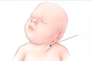
Signs of torticollis in a newborn may appear immediately after birth if the pathology is congenital. Also, the first symptoms may appear within three weeks after the birth of the child. Sometimes the doctor cannot always see the symptoms of torticollis in a newborn, then the first person who can notice the clinical picture of this pathology may be the mother. The most visible symptom is the tilt of the child's head to the sore side. And accordingly, right-sided or left-sided torticollis in newborns will lead to a tilt of the head to one side or the other. Considering that the neck of newborns is quite short and this sign is not always noticeable, then you can see that the child, when lying down, always turns his head to the side. At the same time, his eyes and earlobes are not at the same level. These may be the first signs of torticollis in a baby. Over time, you can see the asymmetry of the baby's face and the muscles on one side may be tense. In a newborn, who has fairly large cheeks, this is easy to notice.
Often, when bathing a child, a mother may notice a symptom of compaction of the affected muscle. This may be when a mother massages a child, she may notice that one muscle is tense or slightly shorter than the other. This may be one of the symptoms that requires further diagnosis.
Congenital torticollis in newborns is also represented by other diseases - this is Klippel-Feil syndrome, Sprengel's disease, and also cervical ribs. All these types of torticollis are congenital, appear in the first month of life and are characterized by curvature of the neck.
Klippel-Feil syndrome is a congenital hereditary disorder that has an autosomal dominant type of inheritance. Classic manifestations of this disease include:
- a short neck in a child, which is quite easy to notice precisely because of the degree of severity of this symptom - after all, the chin can sometimes even touch the shoulder line;
- the hairline at the back is very low;
- when rotating the head there is a pronounced limitation of turns to the side.
Shortening of the neck is usually detected at birth, which directly indicates a diagnosis of torticollis. But sometimes this symptom cannot be noticed, and as the child grows, it is visible how his entire skeleton changes. At the same time, the chest shortens and the lower aperture widens. The shoulders are at different levels and the shoulder blades are very small. Children cannot turn their heads, so they react to any stimuli only by turning their eyes. Then, with age, the next symptom that the mother may notice is that the child does not hold his head, when he should be doing this.
Sprengel's disease is a congenital high position of the scapula. The pathology occurs sporadically, due to delayed development of the scapula and its lowering in the 3rd-4th week of embryonic development. It is characterized by the following main signs:
- Pronounced asymmetry of the neck contours (on the side where the scapula is high, the neck contour is flattened).
- Limited movement in the cervical spine and in the shoulder joint on the affected side.
- High position of the shoulder blade (6-12 cm higher than the opposite shoulder blade).
- Reducing the size of the scapula.
- Rotation of the scapula around the sagittal axis.
- Atrophy of the muscles of the shoulder girdle and shoulder on the affected side.
- Decreased mobility of the scapula, especially with bone fusion.
All these symptoms arise precisely because of the small size of the scapula, so torticollis is considered a secondary symptom.
Acquired torticollis in newborns develops after birth. It can be caused by a bacterial infection or a disease of other organs. And accordingly, there are different types of torticollis:
- Myogenic (due to muscle damage). This can occur with inflammation of the sternocleidomastoid muscle, tumor of this muscle.
- Bone - develops with congenital fractures or dislocations of the vertebrae, as well as with rickets, spondyloarthritis.
- Neurogenic (with pathologies of the nervous system). In children with congenital infantile paralysis with impaired innervation of muscles and their paralysis or paresis.
- Desmo - dermatogenic (with pathology of the skin and ligamentous apparatus). This type of torticollis can develop with large skin scars that can pull the muscles, as well as with eye disease, as a compensatory mechanism.
The most common type of acquired torticollis that can occur in newborns is the so-called Grisel disease. It is characterized by the occurrence of curvature of the neck against the background of inflammatory processes between the I-II cervical vertebrae.
The cause of such torticollis is inflammatory processes of the nasopharynx and ears. In newborns, long-term undiagnosed otitis can quickly lead to a retropharyngeal abscess, which leads to contracture of the muscles near the cervical spine. At first, the child's body temperature rises, which indicates an acute inflammatory disease of the nasopharynx or ear. The sternocleidomastoid muscle on the side of the torticollis is not tense, not shortened. Then the child tilts his head to one side, which can be noticed some time after the symptoms of hyperthermia.
Positional torticollis in newborns is considered one of the simplest variants of the disease. It develops during the first month of life, when the child lies incorrectly in the crib. This can lead to the fact that the muscles on one side can be tense, and on the other, on the contrary, relaxed. This happens if the child sleeps incorrectly or turns all the time to sound or light and lies in this position. When the baby already begins to hold his head, then on one side the muscles are more developed and so the neck is curved in this direction.
Complications and consequences
What is the danger of torticollis in newborns? If it is positional torticollis, then no complications are observed during treatment. But in the case of congenital types of torticollis, the consequences can be very serious:
- secondary changes in bones and muscles in the absence of long-term treatment;
- compensatory scoliosis;
- weakening of pulmonary ventilation with pronounced changes in the skeletal system and disruption of the anatomy of the respiratory tract;
- difficulty breathing and swallowing;
- neurological symptoms (paresis, paralysis, sensory disturbances).
But considering the most unpleasant complication – a cosmetic defect, which is more difficult to correct the more time has passed since its detection, then diagnosis and timely correction are very important.
Diagnostics torticollis in a newborn
Congenital torticollis should be diagnosed in the maternity hospital by a neonatologist. If muscular torticollis begins to manifest itself in the third week of a child's life, it is very important to notice it during this period.
For diagnostics, it is necessary to take into account that in case of pathological birth with an incorrect position of the fetus, especially with breech presentation, clavicle fractures, obstetric brachioplexitis, even in the absence of signs of torticollis in the early stages, this diagnosis cannot be excluded in the future. A characteristic diagnostic sign is that in the 3rd week after birth, at the level of the middle third of the sternocleidomastoid muscle, a dense-elastic formation of varying size appears without signs of inflammation of the soft tissues above the compaction. In the presence of a local compaction of the sternocleidomastoid muscle, it reaches its maximum size and density in the 6th week after birth. Then the compaction gradually decreases, dissolves without a trace and degenerates into a connective tissue cord.
A large group of symptoms are signs that are visible during a simple examination.
The child has a somewhat unusual head position: the head is tilted to the affected side, and it is difficult to turn it to the healthy side. The face is also tilted and the facial muscles may be tense. During examination, an asymmetrical arrangement of the shoulders is noted - on the affected side, the shoulders are higher than on the healthy side. Often there is a change in the contours of the neck - on the side of the torticollis, the contour of the neck is pointed, on the healthy side - flat.
Also, different degrees of facial asymmetry are determined. Due to the growth of the skull, the vertical size of the face decreases and the horizontal size increases. The traction of the sternocleidomastoid muscle deforms the mastoid process; the nasal septum and auditory canal are curved, the upper and lower jaws, and the sinuses are deformed. Also, due to torticollis, compensatory curvature of almost all parts of the spine occurs. Thus, at first the cervical region is curved in the direction opposite to the torticollis. At first, the curvatures are compensatory in nature, but over time, scoliosis develops.
When palpating the sternocleidomastoid muscle, it is sharply shortened, tense, but without signs of inflammation (edema, local and general increase in temperature, soreness, blood changes). Comparative palpation of both sternocleidomastoid muscles (on the side of the torticollis and on the healthy side) is mandatory.
For a more accurate diagnosis, a metric measurement is performed.
When measuring the length of the sternocleidomastoid muscle on the side of the torticollis with a tape measure, its shortening of varying magnitude can be determined. The length of the sternocleidomastoid muscle is measured from the base of the mastoid process to the attachment site of one of the portions of the sternocleidomastoid muscle. The difference in the metric data of the healthy and diseased sternocleidomastoid muscles on the side of the torticollis is the magnitude of the shortening of this muscle. Measuring the angle of the head in the frontal plane also indicates the magnitude of the torticollis. Three degrees of torticollis are distinguished:
- Severe shortening of the sternocleidomastoid muscle - up to 2 cm, head tilt angle - up to 5-8;
- Arterial shortening of the sternocleidomastoid muscle - up to 3 cm, head tilt angle - up to 12;
- Severe shortening of the sternocleidomastoid muscle - more than 3 cm, head tilt angle - more than 12.
How to examine?
Differential diagnosis
Differential diagnosis of different types of torticollis should be carried out with other hereditary diseases that have similar symptoms.
Turner-Shereshevsky syndrome is one of the chromosomal pathologies that are accompanied by symptoms of torticollis. But in addition to this, it is characterized by the following main signs:
- the presence of skin folds on the sides of the neck;
- low or dwarfed proportional growth;
- various forms of chest deformation;
- deafness, cataract;
- exophthalmos, pigmentary degeneration of the retina;
- aortic stenosis, ventricular septal defect.
Often, a geneticist consultation is required for differential diagnosis of torticollis and various congenital diseases.
Who to contact?
Treatment torticollis in a newborn
The approach to treating torticollis depends on the duration of the disease and the degree of expression of changes. Treatment of muscular torticollis in a newborn can be conservative and surgical. Conservative treatment is the use of exercises, physiotherapy and massage.
Massage for torticollis in newborns can be considered one of the first stages of rehabilitation and a very effective method of treatment. How to massage a newborn with torticollis? The massage technique can be as follows:
The baby lies face up on the table, and the mother holds his shoulders in place.
- First exercise. Stroking the sore sternocleidomastoid muscle (at the same time, the head should be tilted back with light movements).
- Second exercise. Using your fingers, massage the affected muscle in a transverse direction.
- Third exercise. The muscle on the affected side is massaged, as if spreading its fibers, the fingers gradually move along the entire neck.
- Exercise 4. Massage of the face and the suprahabial region, which prevents secondary changes in the facial muscles.
- Fifth exercise. One hand is placed on the shoulder joint, the other - in the area of the lower jaw. With slow massaging movements, they try to tilt the head in the opposite direction.
- Exercise 6. The baby's head is grasped with the hand and the face is smoothly turned towards the torticollis, towards the affected sternocleidomastoid muscle. The duration of the massage and the number of movements are gradually increased from 5 to 30 daily.
Exercises for torticollis in newborns should complement massage and can already be performed by the mother independently after several lessons.
A torticollis bandage plays a major role in further consolidation of the results, as it corrects and maintains the results obtained during massage. An orthopedic pillow and orthopedic collar for newborns with torticollis can be used for hypercorrection of the neck position. For children under 6 months, the head can be fixed with a cotton-gauze "donut", a cap. The Shantz collar for newborns with torticollis is also widely used. In this case, the height of the collar on the healthy side is 1-2 centimeters less.
Surgical treatment can be performed if conservative methods are ineffective in children under two years of age. There are also other indications for surgical intervention:
- Forced head position.
- Negative results of active and passive tests.
- Compensatory changes in the skull and spine.
- A sharp shortening and thickening of the sternocleidomastoid muscle.
The operation is performed on children over two years of age. The technique of surgical intervention consists of dissecting the affected muscle. Then, after suturing the wound, a cotton-gauze bandage is applied in the position of hypercorrection of the head. This allows the muscle to form a connective tissue scar already with the correct position of the neck and head. After removing the stitches (7-8 days), a thoraco-cranial plaster cast is applied in the position of hypercorrection, that is, the head should be tilted to the side opposite the operated area, returned to the side of the surgical wound. Fixation with a plaster cast continues for 5-6 weeks. Then the plaster cast is removed and a Shantz-type collar is put on, which is worn for 6 months.
Prevention
Prevention of congenital torticollis consists of the following measures:
- Prevention of pregnancy pathologies (toxicosis, nephropathy, anemia of pregnant women, vitamin deficiencies, infectious diseases).
- Prevention of birth trauma.
- Special care for children at risk (large fetus; breech presentation; transverse fetal position; difficult labor; obstetric clavicle fracture; obstetric brachioplexitis).
- Early diagnosis of pathology.
- Stages of examination of children (maternity hospital, clinic, kindergarten, school).
- Gradual, stage-by-stage treatment of the patient from the moment of diagnosis until the period of completion of growth.
[ 26 ]
Forecast
The prognosis for a child's full recovery is often favorable, with timely treatment tactics. If it is not possible to eliminate the problem with conservative methods, then
the cosmetic effect of surgical treatment is also often favorable. Secondary degenerative changes in the spine sometimes cause severe neurological disorders. Therefore, timely diagnosis and comprehensive treatment are important.
Torticollis in a newborn, when its symptoms appear in this period, is more often muscular. It can be corrected and the child can live a full life after massage and exercise courses. It is only important to start treatment in time and differentiate between different types of torticollis for proper treatment. In this case, the prognosis for recovery is favorable.
[ 27 ]