Medical expert of the article

New publications
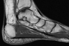
MRI of the ankle: preparation, technique
Last reviewed: 17.10.2021

All iLive content is medically reviewed or fact checked to ensure as much factual accuracy as possible.
We have strict sourcing guidelines and only link to reputable media sites, academic research institutions and, whenever possible, medically peer reviewed studies. Note that the numbers in parentheses ([1], [2], etc.) are clickable links to these studies.
If you feel that any of our content is inaccurate, out-of-date, or otherwise questionable, please select it and press Ctrl + Enter.
Today, magnetic resonance imaging is increasingly being used to diagnose various internal and external injuries and injuries. It is used in various fields of medical practice: from gastroenterology and neurosurgery to traumatology and orthopedics. It gives an opportunity to identify any pathology with high accuracy. Today, the ankle is becoming increasingly important and important. This is a highly informative, non-invasive method that allows to identify the cause and degree of development of degenerative and inflammatory processes in the joint.
Today, rheumatologists and traumatologists are increasingly confronted with injuries and diseases of the ankle, which is explained by the fact that it is subjected to the highest burden. He takes part in all kinds of limb movements, assumes the main load. Withstands the weight of a person. Especially often injuries and diseases develop in women, as they often go on high heels. Athletes, dancers, professional coaches are also most at risk of injury or development of an ankle disease.
What does the ankle show?
An MRI can show a lot to a specialist. With this method, you can visualize the basic structure of the joint, so you can quickly get the right diagnosis and choose the necessary treatment. You can diagnose pathological conditions, identify trauma. Gives a lot of useful information in the diagnosis of bones, tendons, ligaments and bones of the joint being examined. It is also possible to identify tumors of any genesis and stage, arthritis, bleeding and bruises in a timely manner.
The advantage of the method is the ability to identify chronic hematomas and lesions, which is widely used in forensic medicine in carrying out surveys.
The method can show lesions of a different nature in the ankle, Achilles tendon. It is the tendons and ligaments located here that provide the flexibility and mobility of the joint, enabling it to carry out the entire range of movements characteristic of it.
With the help of MRI it is possible to identify tears and complete ruptures of ligaments and tendons of the joint, their stretching, mechanical damage, inflammatory process. It makes it possible to reveal the slightest changes in the structure of the cartilaginous tissue. Also, various thinning, involution, degenerative processes are visualized.
During the procedure, a good visualization of the bones of the ankle and foot is ensured. You can see even the talus and heel bone, which are almost impossible to explore by other methods. This is practically the only method of determining fractures of these bones. You can also detect bruises, dislocations, and signs of osteoarthritis, arthritis, and osteoporosis.
The method is very informative in preparation for surgery, as it makes it possible to identify the presence and localization of tumors, visualizes the accumulation of blood and exudate in soft tissues, around the joint, or inside it. It allows to assess the state of the distal sections of the tibial and fibular bones, as well as the muscles of the foot. You can additionally enter a contrast, which will give an opportunity to explore the structure of the ankle in detail and determine even minimal morphological changes. You can visualize dystrophic, degenerative, inflammatory processes.
Indications for the procedure
The procedure is appointed, if necessary, to examine the ankle joint, in particular, with injuries of tendons, ligaments, cartilage. The procedure is informative if necessary to detect a fracture, dislocation. This is practically the only method that makes it possible to identify tumors in the early stages of their development. Can be visualized as a tumor of soft tissues, and bones, joints.
Assign for the diagnosis of infectious and inflammatory processes, necrosis. It makes it possible to identify false joints and unconsolidated fractures, such diseases as arthritis, arthrosis, tendonitis, tendonosis.
Assigned in the presence of congenital anomalies and pathologies, with the development of pain, swelling, redness in the ankle. It is used as an additional research method with insufficient information content of other methods. For example, to clarify the diagnosis in the case if an x-ray was detected, but the pathology was not completely differentiated. It is prescribed with a decrease in the volume of movements in the joint region, an unclear genesis of pain in the joint. Must be used in preparation for operations.
 [
[Preparation
Before the procedure, the patient must take off his clothes and be in a special disposable clothing. It is allowed to stay in your clothes only if it is free-cut and does not contain metal parts and inserts.
In the protocols of the study, the mechanism of catering before and after the procedure is not prescribed. Based on practice, doctors recommend refraining from eating a few hours before the study. This is especially true if the study is planned with contrast. It is also important to inform before the procedure starts about the presence of allergic reactions, intolerance of some components. Also be sure to inform the doctor about the presence of bronchial asthma.
The contrast medium used contains a metallic component - gadolinium. It has practically no side effects and does not entail complications. Nevertheless, people with severe physical illnesses, heart and kidney pathologies should not use it. At least, the presence of such concomitant diseases must necessarily be reported in the archa in advance.
It is important to get information about pregnancy in advance. Therefore, if a woman doubts, in preparation for the study it is necessary to take an analysis for pregnancy. It will be enough to analyze for hCG.
Before the procedure, the patient is explained what and for what purpose will be investigated which procedures will be applied. It is important to inform the patient about the expected results, risks, consequences of the procedure. Claustrophobia recommends the use of open devices. For children, preliminary sedation is mandatory, which will enable the child to lie quietly and motionless, thus avoiding injuries during the procedure.
It is necessary to remove and remove all objects that contain metal. You need to make sure that all jewelry, watches, lined business cards, credit cards are removed. Also removed hearing aids, dentures, piercings. Lay out handles, pocket knives, glasses and any other objects.
Technique of the mRI of the ankle
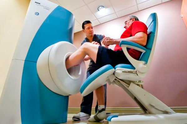
Traditionally, a closed-type MRI device is always used. O has the form of a large cylindrical tube. Which is surrounded by a magnet. During the procedure, the patient is placed on a movable table. Which moves towards the center of the magnet.
There are also open-type MRIs, but they are less informative because the magnet does not completely surround the patient. From the sides it remains without a magnetic part. This method is used only if the person has claustrophobia, or at a large weight.
When examining the ankle joint, the coil is placed directly on the test joint. The patient should be in a lying position, remain motionless. On average, the procedure lasts from 30 to 40 minutes. If a study is done with contrast, the procedure lasts longer.
The procedure is painless. Some patients note the emergence of specific sensations in the area where the study is conducted. It can be tingling, vibration is warm, light burning. Each impression is individual. This is normal, you should not worry. This is how the individual reaction of tissues to the magnetic effect manifests itself.
During the study, the patient is alone in the apparatus room, but between the doctor and the patient eats a two-way audio communication. The doctor sees the patient. After the procedure, no adaptation is required.
Today, it is possible to carry out an ankle ankle with small devices that do not require the full placement of a person in the camera. Only the necessary joint is examined. The image is of a sufficiently high quality.
MRI of the ankle ligaments
Often there is a need for ankle ligament research. The most effective method for this is precisely MRI. It provides an opportunity to comprehensively examine the Achilles tendon, assess its condition, and identify possible pathologies. Used to detect tears and tears. Sometimes other ligaments are examined, if they cause pain or suspicion of a pathological process. Often the study is subject to the deltoid ligament, which stabilizes the joint. What kind of ligament is damaged, can often be detected only by the results of an MRI scan.
Contraindications to the procedure
The MRI procedure can not be performed if the patient has different implants, implanted electronic devices, with tattoos containing iron, metal impurities.
MRI is contraindicated in the presence of pacemakers, endoprostheses, defibrillators. It can not be performed with artificial heart valves, with some types of clips that are used for aneurysms of the brain, with metal spirals that are located within the blood vessels.
Contraindications include implanted nerve stimulators, metal pumps, pins, screws, plates, surgical staples. Also, the procedure is not carried out if there are any metal parts in the human body, for example, bullets, a fragment. This is due to the fact that the magnetic field will attract metal to itself, and will move it, which can lead to tissue damage, rupture of blood vessels.
Complications after the procedure
The procedure has no complications. Exceptions are cases of non-compliance with safety rules. If the procedure is performed in the presence of contraindications, serious complications, up to a lethal outcome, are possible.
This is due to the natural effect of magnetic particles: if there are metal elements in the human body, or implants, they are attracted by a magnetic field. This can lead to their displacement, breakage. As a result, tissue and vascular damage, bleeding, irreversible effects can occur.
At present, nephrogenic systemic fibrosis is recognized as a possible complication after the administration of a large amount of contrast medium. But this effect is extremely rare. It often happens in patients with renal insufficiency or other serious disorders of the structure and function of the kidneys.
Consequences after the procedure
The procedure is absolutely painless and harmless and has no consequences. Adaptation after the procedure is not required. A person can immediately go on vacation or do business. In rare cases, the development of an allergic reaction to injected contrast agents. This is observed in the event that a person suffers from an allergy and is not warned in advance. Claustrophobia may occur if a person suffers from this disease. Nervous seizures and seizures occur in people with serious neurological disorders and severe mental conditions.
Reviews
If you analyze reviews, you can see both positive and negative feedback. As noted by many specialists who use this method in their diagnostic practice, MRI is a highly informative accurate method. The big plus is that it is non-invasive and does not require any preliminary preparation. Provides a high level of visualization and does not allow the use of ionizing radiation.
It is a valuable method for diagnosing a wide range of conditions, including inflammation, injury, and trauma. It is used almost always before surgical interventions. It allows the surgeon to obtain the most accurate information and determine the scope of surgical intervention. It is possible to diagnose complex fractures, even in those cases when the x-ray does not yield any results. Also, it is possible to detect those anomalies that are not visible when examined by other methods.
However, they also note the risks associated with this procedure. Sometimes sedation is required, because a person may have claustrophobia, or he can not stand the time of the procedure motionless. Sedation is also applied to children. Sometimes a person is too nervous, the device seems daunting, so you have to inject sedatives. There is always a risk of excessive sedation.
Despite the fact that the magnetic field itself does not act negatively on a person, implanted devices or metal elements in the human body can lead to serious damage. Also, there is always a risk of developing an allergic reaction, especially when using a contrast medium. But usually such reactions are quickly stopped by the introduction of antiallergic drugs. There is always a risk of developing an attack of claustrophobia when using a closed-type device.
Patients characterize the anterior malleolus as a painless procedure. Some are embarrassed by the need to immerse in the apparatus, which causes concern. After the procedure, there is no discomfort, well-being.