Medical expert of the article

New publications
Hypoplasia of enamel of permanent and milk teeth
Last reviewed: 23.04.2024

All iLive content is medically reviewed or fact checked to ensure as much factual accuracy as possible.
We have strict sourcing guidelines and only link to reputable media sites, academic research institutions and, whenever possible, medically peer reviewed studies. Note that the numbers in parentheses ([1], [2], etc.) are clickable links to these studies.
If you feel that any of our content is inaccurate, out-of-date, or otherwise questionable, please select it and press Ctrl + Enter.
The pathology of the structure or mineral composition of the dental tissue (partial or complete absence of it), which develops due to a malfunction that arose even in the period of their formation, is a hypoplasia of the teeth. This disease is of a fairly common nature.
In one form or another, it affects about 30% of the population. More often this problem is subject to permanent teeth, less - their dairy predecessors. The most severe manifestation of the disease is the "underdevelopment" of dentin, and its extreme manifestation is the complete absence of enamel or tooth as a whole.
Doctors state that the number of sick children and adolescents significantly exceeds the percentage of adults who suffer from this disease. Often the most vulnerable place for hypoplasia of teeth becomes enamel: it is less durable and the thickness of the enamel coating layer is lower than the norm presupposes. The presence of a given disease in a person often speaks of a serious enough pathology of metabolic and protein processes in the body, thus being a separate disease and, at the same time, a symptom of a deeper pathology affecting the human body.
Causes of tooth hypoplasia
To successfully combat the disease, it is necessary to thoroughly know the causes of its appearance. One of the main reasons for the development of pathology is associated with disorders that have arisen in the system of protein and mineral metabolism metabolism (the destructive activity of this disease is irreversible). What are the causes of dental hypoplasia:
- This failure may occur on the basis of Rh-conflict, which develops in the mother with its fetus.
- If the future mother during pregnancy, especially in her first trimester, suffered an infectious disease.
- If the pregnancy was severe, with severe toxemia.
- The child was not born full, prematurely.
- The kid was injured during childbirth.
- Pathology of child development during the infants: rickets ...
- The kid does not get the necessary amount of food - dystrophy.
- Diseases of the gastrointestinal tract.
- Violation of metabolic processes. In particular, calcium.
- Present somatic diseases in the child.
- Disorders of brain activity, which manifested itself at the age of six months to a year ...
- Infectious lesions.
- Mechanical injuries of the maxillofacial region.
Symptoms of tooth hypoplasia
There is a symptomatology of this disease, and the specifics of their manifestations largely depends on the severity and complexity of the factors and diseases that the patient had to endure.
There are several types of disease, in which the symptoms of dental hypoplasia slightly differ.
Systemic hypoplasia (pathology affects all the teeth of the patient's oral cavity):
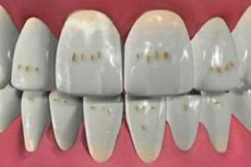
- The color deviation (in comparison with the norm) of the tooth enamel. The presence of only this sign can be attributed to the manifestation of mild degree of hypoplasia of the teeth. The spots are distinguished by a clear outline, having a white, less often yellow, shade. These spots are located on the front wall of the tooth. Painful discomfort does not cause such a deviation. At the same time, the surface of the enamel does not change its texture (as smooth and glare as at the norm).
- Insufficient thickness of the enamel layer or its complete absence.
- Underdevelopment of the layer covering the tooth.
- Individual cases of this type of disease can be called "tetracycline" teeth. They are very different in color from classical teeth. Their color was changed by the action of such a drug as tetracycline, which was actively taken throughout the period of formation of Mom's teeth, as well as from the lack of necessary minerals in this period.
Local hypoplasia (pathology affected one or two teeth):
- At this type of disease, pathology affects not only the enamel, but also the deepest layers - the rudiments, from which the permanent teeth are subsequently cut. For example. They may be prone to inflammation that occurs in the jaw (due to infection), or arose after the resulting mechanical trauma.
- Defect in the development of tooth enamel. This is quite a serious case of illness. The tooth suffered more significant damage. On its surface, structural defects (bands and minor dents) are seen. Breakdown in the integrity of the surface of the enamel is not observed. Painful symptoms do not appear. The patient is more concerned about the aesthetic side of his smile.
- Aplasia is the most common but the most severe form of hypoplasia of the teeth. This pathology is only congenital. The main and most unpleasant symptom is when the patient's teeth completely or partially lack the enamel material. In this case, psychological pain is also added to the psychological discomfort - the reaction of open, unprotected areas of the dental tissue to external stimuli: touch, temperature fluctuations, various physical and chemical substances.
- There are cases of underdevelopment of dentin, which leads to deformation of the tooth itself (it can take quite bizarre forms).
Hypoplasia of tooth enamel
It will be absolutely incorrect to state that hypoplasia of the tooth enamel depends only on the quantitative and qualitative "filling" of the human body with mineral substances. And with demineralization - we get a hypoplasia of teeth. If this were so, then there would not be such a scale of the disease and problems with its treatment. The scale of the disease is increasing every year, because the adverse effects on the rudiments of the teeth begin in the womb, at the time of the laying of genetic information. And as a result - more than half of the children suffer to a greater or lesser degree on the hypoplasia of the teeth. Scientifically proved that aplasia of tooth enamel is much more common in children whose mothers during pregnancy have suffered acute infectious and respiratory diseases, toxoplasmosis, or an extreme form of toxicosis. Hypoplasia of tooth enamel a baby can receive and in case of prematurity, as a result of trauma received during birth, as well as with poor diet, severe forms of diseases transferred to the infants.
The enamel pathology is found in both dairy and permanent teeth, with the latter accounting for the highest percentage of diseases, leading to an increased risk, and further, to get other diseases.
Hypoplasia of teeth in children
Hypoplasia of teeth in children is a common pathology. She suffers in an easy or more difficult degree every second child.
If the pathology of the milk teeth with its "roots" goes to the violations that the fetus received in the womb, the hypoplasia of the permanent teeth is a disease acquired by the child (alone) after birth, it is provoked by malfunctions that occurred in the child's exchange system from the moment, when he reached the age of six. Since up to the year the child is sick much more than the intrauterine pathologies that occur, then, naturally, the hypoplasia of permanent teeth (and its systemic form) is diagnosed in most cases revealed.
The pathology of permanent teeth is formed under the influence of diseases and their complications, which can be overtaxed at the age of 0.5-1.5 years. These are diseases such as rickets, acute infection, dystrophy, diseases of the gastrointestinal tract, disorders of brain functions. The localization of such spots directly depends on the age of the child in which he suffered this disease, and the depth of the lesion depends on the severity of the course of the disease.
It is noticed that if the youngster was sick at the age of 5-6 months, the central incisors (cutting edge) and the thighs of the 6th teeth that form during this period will be affected by the enamel. If the disease has fallen for a period of 8 to 9 months, then the affected will be fangs and second incisors. Since the timing of the laying of teeth are different, then the sites of hypoplasia are located in different areas of the teeth. But if the disease is prolonged or has passed into a chronic condition, the baby can get an aplasia - complete absence of enamel on the surface of the teeth.
The hummocky texture of the enamel can speak of a prolonged, periodic recurrence of the course of the disease, and its severity affects the depth of the lesion of hard dental tissue. That is, an easy disease can only result in the appearance of small spots on the tooth, whereas a serious infectious disease can lead to the complete absence of the enamel layer on the tooth.
Hypoplasia of infant teeth
Perennial medical studies have shown that hypoplasia of the infant teeth is due to those harmful factors that affected the unborn child in the womb. Especially dangerous in the development of pathology are the infectious diseases transferred by the mother during pregnancy, acute respiratory infections, rhesus - conflict with her baby, strong manifestations of toxicosis ...
Forms of hypoplasia of teeth
To date, medicine does not have a specific and universally accepted classification of pathological forms. And yet, let not officially, but this classification of the form of dental hypoplasia still exists.
- Stage of the spot. This form is due to the appearance on the chewing, oral and vestibular areas of the dentition of a rounded configuration of spots of milky white (somewhat less often yellowish) color. It is interesting that the teeth are affected symmetrically, affecting the same name. Usually the configuration of the spot is not blurred, clearly delineated, not lost its luster. The gloss and the smooth surface of the spot may indicate that the affected tooth was exposed (not so much) to external, negative factors, and this action proceeded for a short time. Although, all the same, and reflected on the texture of the enamel, its structural components and the amount of minerals that are included in this conglomerate.
If the affected area has a rough texture and a dull color - this indicates that the enamel was subjected to destructive influence at a time when the main stages of formation were already passed. Such changes affect only the surface areas, while the amount of the enamel layer remains unchanged.
The patient does not feel pain, there is no increased sensitivity of the tooth to temperature, mechanical and chemical stimuli.
Hypoplasia of the teeth, in any form of its manifestation, will not disappear on its own, mandatory medical intervention is necessary.
- Cup-shaped (erosive) shape. Round-oval cup-like defects, differing in different sizes (depth and diameter). This form can be called a steam room. Erosion, as a rule, is located on symmetrical (the same name) tooth surfaces, while showing the same shape and size. The closer to the bottom (bottom) of the bowl, the enamel becomes thinner. In this case, the penetration of the dentin from the deeper layers gives the spot a yellowish shade. A more radical case may also appear - aplasia of the enamel. That is, at the bottom of the hole, the layer of enamel can be completely absent. All the surface of the well is smooth.
- Borozdchataya form. When visual examination in the vestibular region of the affected tooth, one or several furrows are clearly visible. Most often they are located parallel to each other and the cutting edge. The depth of the rut is different and depends on the severity of the lesion. The thickness of the layer of enamel in the hollow of the furrow can vary from the norm to its complete absence (the dentin is clearly visible). The lesions occur symmetrically, on the teeth of the same name. This form of hypoplasia of the teeth is perfectly visible on the X-ray and can be detected even at the stage of eruption. The picture clearly shows the clarified furrows with well-defined edges. They are arranged horizontally.
- Linear (wavy) shape. This variety can be characterized as a set of multiple horizontally located grooves that are localized in the vestibular area of the tooth surface. This fact also makes the structure of the enamel wavy.
- Aplastigeskaya form. Severe case of hypoplasia of the teeth. With it, the surface of the enamel is completely absent on the hard tissues of the tooth, or partially present in small areas. It occurs when abnormal violations of amelogenesis.
- Mixed form of dental hypoplasia. Already the name speaks for itself - a combination of several forms. More often, one person may have both a spotted and a cup-shaped form of this disease. This somewhat complicates the diagnosis.
 [6]
[6]
Specific form of hypoplasia of the teeth
To this form carry (names on surnames of scientists who described them in detail):
- Getschinson's teeth. These are usually incisors, which can be located on both the upper and lower jaws. The shape of the tooth is barrel-shaped, the cutting edge is a crescent.
- The teeth of Pfluger. The shape is very similar to the Getschinson's tooth, but in this case there is no crescent edge.
- Teeth of Fournier. Most often these are permanent molars, cut first. The shape of the tooth is conical, with slightly protuberant tubercles. This form is often associated with congenital pathology - intrauterine syphilis.
Diagnosis of tooth hypoplasia
Diagnosis of tooth hypoplasia is performed by a dentist on the basis of a visual examination of the patient. One of the main difficulties is to differentiate this disease from tooth decay of the tooth, although often enough these two diseases go hand in hand.
Caries usually gives a solitary spot on the surface of the enamel in the tooth's neck, whereas the hypoplasia of the teeth is manifested, more often, by a lot of whitish spots that are dispersed in different parts of the tooth.
A 2% solution of blue methylene can become a litmus in diagnosing the disease in question. In case of caries disease, the stain is stained, and in the case of tooth hypoplasia, discoloration does not occur. Also, with caries, the surface of the spot becomes rough, and the surface remains smooth for the disease under consideration.
What do need to examine?
Who to contact?
Treatment of dental hypoplasia
If the disease is diagnosed at an easy degree, the spots are small and slightly noticeable, in which case the treatment of hypoplasia of the teeth is not carried out. If the symptoms of this disease perfectly differentiate with a smile or conversation, especially if the deep layers of dental tissues are affected, treatment is clearly necessary. And you need to hold it as soon as possible. Procrastination can provoke unpleasant consequences:
Complete loss of the affected tooth or all teeth.
- The tooth edge is erased much faster than normal.
- Destruction of hard tooth tissues.
- The bite defect develops, which in the future can lead to gastrointestinal diseases.
The protocol of treatment is somewhat different for different forms of the disease. If the disease has not acquired a global scale, then the basis of treatment can be teeth whitening, with its severe manifestations - this is a filling. If the shape of the tooth is defective, the dentist is going to grind the irregularities that are possible with pathology. If necessary, the dentist will not only fill the affected teeth, but also their prosthetics.
Bleaching can be carried out under the supervision of a dentist both professionally and at home.
Whitening at home will cost the patient cheaper and more comfortable, but will last longer than with a clinical approach to the problem.
The most popular method of bleaching to date, you can call a special device (kappa). It is made individually for a specific patient on the recommendation of a doctor. The device is filled with a special whitening gel and worn for 3 to 10 hours at home. The procedure is long enough, but more effective than with whitening pastes and chewing gums.
- Bleaching gel Plus White Whitening Booster
- Before applying the whitening gel to the tray, it must be rinsed and dried.
- To sanitize the oral cavity with toothpaste, brushing your teeth (preferably using a fluorine-containing compound).
- With a special syringe - container, apply the gel evenly to the tray.
- It is necessary to dress it on the dentition, pressing it well. Remove excess gel with a napkin.
- The time of the procedure depends on the percentage concentration of the drug.
- 10% - for the night or six - ten hours.
- 15% - four - six hours.
- 20% - two - four hours.
- 35% - half an hour.
- After using the kappa rinse, rinse thoroughly with warm water mouth.
This gel was specially developed for home use.
- Colgate Simply White Night Gel
- This gel is applied once a day and applied before going to bed on the teeth, previously cleaned with toothpaste. It must be remembered that the whitening gel is applied to the dry enamel.
- Gel, with the help of a complete brush, carefully applied to each tooth separately. One drop of the brush into the bottle is enough for three teeth.
- After application, the preparation is quickly absorbed by dental material, so there is no need to dry or rinse the oral cavity with teeth.
- After this procedure, do not eat or drink for 15 minutes.
- Three to five days is enough to make the result obvious.
- With prolonged use of whitening gel, tooth enamel can brighten by three to four tones.
- It is also necessary to take precautions: make sure that the product does not get into the eyes (in case of contact, rinse immediately with clean water), this medication should not be used by children under 12 years of age.
- Store in a cool place.
- Whitening effect does not take about a year.
- Gel ROCS Pro "Oxygen bleaching"
An excellent whitening effect of this drug due to the properties of its active oxygen. This medical device perfectly suppresses the inflammatory process in the oral cavity, thereby removing the unpleasant odor that comes from the mouth.
The gel perfectly penetrates into the deeper layers of enamel and dentin, thereby brightening the tooth by two to three tones. The optimal treatment course is four weeks. A low abrasive index makes it possible to use this gel when cleaning with both classical and electric brushes. But this is also a minus of this medication, as it does not have a polishing property, which reduces the effectiveness of removing surface shades.
Any bleaching gels should be used with caution and not for a long time, as during the treatment procedures with the use of bleaching agents, the enamel becomes thinner, the sensitivity of the teeth increases. The frequency of using such pastes for preventive purposes should not exceed two times a week, and in medical - according to the prescribing physician.
The proposed advertising for bleaching chewing gum can only be conditionally called an effective bleaching agent.
- Professional approach to whitening. This procedure is performed only in the conditions of a specialized dental clinic.
- Airflow is a professional in whitening. To date, this is one of the most popular professional methods of whitening in dentistry. The procedure allows you to lighten the enamel of the teeth by several tones. This is one of the fastest and safest ways to achieve the desired result.
In the process of processing, not only clarification of the enamel occurs, but also the tooth is cleaned of tartar and plaque, which give the tooth a shade of yellowness and is a breeding ground for pathogenic bacteria. This is what allows you to achieve the effect of whitening, bringing the enamel closer to natural color.
This procedure can be called a litmus test in the diagnosis of dental hypoplasia. Approaching the shade to their natural color, the dentist can more closely examine the surface of the enamel, identify the foci of lesions and decide on the need for further treatment.
The procedure of clarification with the help of the Airflow method is carried out in the presence of a special device that creates an overpressure and in this environment, with the help of specialized pastes, gels, therapeutic mixtures, the tooth surface is treated.
The principle of the device: At the tip of the nozzle, which is inserted into the mouth, sodium bicarbonate powder is mixed with the aqueous suspension and pressurized air. The result of this procedure is the removal of plaque, bacteria, minor dental deposits, light grinding of the surface. The procedure does not take much time, it does not bear pain. Thus, the patient will spend a few minutes in the clinic and will receive the same result as in a few weeks at home.
To consolidate the success and prolong the result, a special protective varnish is applied to the treated surface.
During the treatment, the tooth loses the cuticle. Therefore, two to three hours after the procedure, it is not allowed to drink coffee and tea, smoke, consume foods that have enzymes that color them. The formation of a new cuticle occurs from the saliva through this time interval.
The method of bleaching Airflow is safe, but still has some limitations:
- A number of periodontal diseases.
- Hypersensitivity to the taste of citrus.
- Pregnancy.
- Breast-feeding.
- Children of young and early adolescence.
- Asthma.
- Chronic form of bronchitis.
- Diseases that involve a salt-free diet.
- Teeth whitening with laser. This method is quite in demand today in domestic and world dentistry. He does not cause any discomfort to the patient, while not taking much time. But the effect of the procedure can last for several years.
Bleaching is done in a clinical setting using a medical device that produces a Whitening Accelerator diodes laser beam. Cleansing preparations are special gels or pastes. Contraindications for use are the same as in the previous case. But if the use of the Airflow method, seals are not a contraindication, then with laser correction, seals on the facade of the dentition can lead to uneven shades.
- Photo whitening. It is carried out strictly in the conditions of the dental clinic. Activation of a special whitening paste or gel is due to specialized medical equipment - photolamp. Resistance of the obtained result reaches one to three years.
In parallel with this, the patient necessarily receives drugs that will help restore the mineral composition of the tooth enamel.
- Remodent
For rinsing, which lasts 3 to 5 minutes, prepare a 3% solution (in 100ml of boiled water, dissolve 3 g of the drug). For therapeutic purposes rinsing is carried out four times a week, but not more than 40 rinses. Prophylactically - for ten months, from two to eight rinses per month. A side effect may be an allergic reaction to the drug. After rinsing for two hours, do not recommend eating and drinking.
- Calcium gluconate solution
This medical device slowly (for 2 to 3 minutes) is administered 5 to 10 ml intramuscularly or intravenously, preheating to body temperature. The schedule of injections is one to two days, depending on the complexity of the disease.
At admission, side abnormalities may appear: nausea, up to vomitive reflexes, as well as bradycardia and diarrhea. Medication can not be taken to people suffering from blood diseases: thrombosis, increased blood coagulability.
Prevention of hypoplasia of teeth
Prevention of hypoplasia of teeth includes a set of measures that can prevent diseases, a complication of which can be a systemic metabolic disorder in humans. Therefore, it is necessary to treat any diseases in a timely manner without transferring it to the chronic level.
It must be remembered that the teeth are formed in the future little man still in the womb (milk teeth), therefore, to prevent the development of hypoplasia of teeth, nutrition should be balanced. This is especially true for both the future mother and newborn, because the permanent teeth develop in the first months after birth.
In the diet of mother and child, as well as any person, there must be products:
- With a high content of fluoride and calcium: cottage cheese, milk, cheese and others.
- Vitamin D. In the form of tablets or you need to take adequate time to sunbathe.
- Vitamin C. These are citrus fruits, Brussels sprouts and broccoli, green onions, spinach, currants, rosehips ...
- Vitamin A. It is liver, garlic, sea kale, seafood, butter, broccoli and others.
- B vitamins are nuts, pork, poultry, cereals, legumes (especially lentils), mushrooms, fish and others.
As the baby grows, the consistency of the food should change, since with malnutrition, diseases of the gastrointestinal tract can develop, which also can lead to malfunctions in metabolic processes. A child of 0 - 3 months old age should eat only liquid food, from 4 to 6 months - homogeneous liquid, from half a year to 9 months - by grinded food, the last two months (up to a year) - the products should be chopped, from year to year and a half - and already from three years - a full-fledged classic dish.
To prevent the development of dental hypoplasia, the child, under the supervision of an adult (and an adult on his own), must maintain oral hygiene: twice a day with a properly selected toothpaste and brush to brush your teeth. After each meal, rinse your mouth, eliminating the remnants of the meal.
During the meal, you must thoroughly chew the food. And also to visit a dentist on time, preferably for preventive purposes.
Introduce in your diet meals that will strengthen your gums and teeth.
For example:
- Spring salad: Thoroughly wash and chop the leaves of the young nettle and dandelion, add feathers of spring onions. Salad dress with vegetable oil. To improve the taste, you can introduce a boiled egg and lightly salt.
- French salad. Take sprouted wheat and oat flakes (enough for two tablespoons of each component), grind. This gruel is poured for an hour with six tablespoons of boiled water. After the product is infused, add 3 tbsp. Spoons of warm boiled milk, 1 tbsp. A spoonful of sugar, the juice of one lemon and one grated apple with a peel on a large grater.
- Salad is refreshing. Remove one large apple from seeds and peel, cut into pieces, 250g of cheese into small cubes. Celery, pre-cooked in salted water, chop. All carefully mix, season with herbs (parsley and dill), pour 4 tbsp. Spoons of vegetable oil and 1 tbsp. Spoon of vinegar.
- Vegetable salad. Grind carrots and celery (1-2 small root crops), pepper, fresh cucumbers and leeks, add 300 g of canned or boiled corn. Salad season with vegetable oil or mayonnaise.
This list of dishes to prevent the occurrence of hypoplasia of the teeth, you can continue indefinitely. If desired, they are not difficult to find in the specialized literature, in the Internet space or, having shown imagination, to invent yourself.
Prognosis of tooth hypoplasia
In many respects, the prognosis of dental hypoplasia depends on the form and type of the disease, which is revealed in the patient. If slight local hypoplasia of the teeth is observed, then the defects are stable and do not cause any complications. In this case, treatment is not required, it is only necessary to adhere to the norm of nutrition and hygiene. If the dentist observes systemic dental hypoplasia, he makes up a protocol of treatment, which is carried out. The level of development of modern dental science allows to solve this problem with honor. Even if the patient suffers the most severe form of the disease - aplasia, the way out of this situation also exists - partial or full prosthetics of the oral cavity.
Such an easy and such a complex disease - hypoplasia of the teeth. She is to a different extent sick third of the world population and every second child under 12 years old. But the steps that dentistry has done and is doing in recent years are encouraging. The main thing is not to discount those preventive measures that will save you and your baby from this unpleasant illness. But if the trouble has come - do not delay with a hike to the dentist. This specialist will do everything to make your smile radiant and snow-white.