
All iLive content is medically reviewed or fact checked to ensure as much factual accuracy as possible.
We have strict sourcing guidelines and only link to reputable media sites, academic research institutions and, whenever possible, medically peer reviewed studies. Note that the numbers in parentheses ([1], [2], etc.) are clickable links to these studies.
If you feel that any of our content is inaccurate, out-of-date, or otherwise questionable, please select it and press Ctrl + Enter.
Diagnosis of breast cancer
Medical expert of the article

Last reviewed: 06.07.2025
Breast cancer diagnostics is a set of methods aimed at identifying malignant tumors. The effectiveness of treatment and prognosis of recovery depend on early detection of this pathology. Let's consider the main procedures used to identify malignant lesions.
Breast cancer is the most common form of tumor in women, accounting for 19% of all malignant neoplasms. The risk of malignant neoplasms increases in direct proportion to age. The highest percentage of fatal outcomes occurs in women aged 40-55. In men, breast cancer is diagnosed very rarely. There are a number of risk factors that can cause malignant neoplasms. The main factors are: menopause, fibrocystic mastopathy, no childbirth or childbirth after 30 years, family history, etc.
The localization of the tumor in the mammary gland may be different. With equal frequency, both the left and right glands are affected, and in 25% of cases, bilateral cancerous lesions are observed. In this case, the node in the second gland can be both an independent tumor and a metastasis. Most often, tumors appear in the upper-outer square or near the armpit. The main symptoms of the disease are manifested as compaction and retraction of the nipple, bloody discharge from the breast, pain.
In addition to the classic clinical picture, the following forms of the disease can be identified during the diagnostic process:
- Mastitis-like
A sharp increase in the mammary gland, swelling, pain. The skin becomes hot to the touch, turns red. To identify mastitis-like cancer, a differential diagnosis is made with acute mastitis.
- Erysipelas-like form
During an external examination, the first thing that is noticed is red skin, and the redness extends beyond the chest. In addition to the redness of the skin, the patient has a high temperature. Diagnosis should be made by an experienced doctor, since this form of malignant neoplasm is often confused with ordinary erysipelas.
- Armored
It appears due to cancer infiltration through skin crevices and lymphatic vessels. The skin thickens and becomes bumpy. A thick skin shell forms on the chest, which can cover one or both glands. The disease has the highest degree of malignancy.
- Paget's cancer
It is a flat lesion of the nipples and areolas. With early diagnosis, the main sign of the lesion is wet and flaky nipples, which can be mistaken for eczema. With further development, cancer grows into the gland ducts and forms a node with metastatic lesions of the lymph nodes.
The doctor's task is to identify all the typical symptoms of cancer. The examination is carried out not only on the affected, but also on the healthy breast, in order to identify the presence of metastases. Palpation of the supraclavicular and axillary cavities is mandatory. If the symptoms of a malignant neoplasm are clearly expressed, then diagnosis is not difficult. But if the disease is in the initial stages, small in size or deep in the tumor, additional diagnostics are carried out.
As additional diagnostics, non-contrast mammography, radiography, aspiration biopsy, puncture with cytological examination are used. Partial excision of the tumor together with surrounding tissues and histological examination are possible. If the presence of cancer is confirmed, the operation is expanded to radical. To determine the extent of spread of the malignant tumor in the body, the patient undergoes skeletal scintigraphy,liver ultrasound, bone radiography and lung X-ray.
Early diagnosis of breast cancer
Early diagnosis of breast cancer is a comprehensive approach that consists of many methods used in both medicine and oncology. The main goals of early diagnosis are:
- Detection of cancer at an early stage (this is the period when successful treatment can be carried out).
- The doctor's choice of an effective and ideally suitable treatment method.
- Evaluation of therapy results.
The diagnosis should answer questions such as: what type of tumor (invasive or non-invasive), are there metastases in neighboring lymph nodes, and if so, how large is the lesion.
Early diagnostics is divided into primary and clarifying:
- Primary diagnostics
The examination is called screening. Its main task is to identify primary changes in the mammary gland. This is a self-examination of the breast, palpation of the mammary glands, examination by a surgeon, mammologist, oncologist and endocrinologist. Primary diagnostics is carried out in women without obvious signs of changes in the breast. Examinations should be regular, since their goal is early detection of malignant neoplasms.
- Clarifying examinations
In this case, methods are used that allow a targeted search for changes in the mammary glands. Diagnostics makes it possible to clarify the nature, prevalence and character of changes. Examinations are carried out throughout the treatment to monitor its effectiveness. The main diagnostic methods in this category are: MRI, ultrasound, CT, biopsy and others.
Early diagnostics of breast cancer is performed by a doctor at each visit to a women's clinic. Standard diagnostics involve palpation of the organ to determine the presence of swelling and painful lumps. This examination is explained by the fact that a malignant neoplasm of the breast most often manifests itself as a small swelling, which in 90% of cases the woman discovers herself. During the examination, the examination is carried out in a lighted room, in a vertical and horizontal position, with raised and lowered arms.
During the examination, the doctor pays attention to a number of factors that may indicate the presence of pathology: swelling or hardening of the breast (nipples), redness or swelling of the skin, asymmetry, changes in the shape and position of the nipple. Deformation of the areola, discharge from the nipples, retraction of the skin on the chest, peeling of the breast, tumor seals in the armpit, swelling of the shoulder, pain and discomfort in the chest also indicate a pathological process.
Very often, obvious signs of cancer are diagnosed at late stages, when the tumor takes on an advanced form. In this case, a dense painful neoplasm grows into the chest wall, which leads to immobilization of the breast. Due to the tumor growing into the skin, the mammary gland ulcerates, becomes deformed, and the nipple is drawn in. Bloody discharge may appear from the nipple. If the tumor grows into the lymph nodes, this leads to an increase in the axillary lymph nodes, which causes pain and discomfort.
 [ 4 ]
[ 4 ]
Differential diagnosis of breast cancer
Differential diagnostics of breast cancer are examinations that allow to exclude the disease by certain factors and symptoms, which will ultimately allow to diagnose the only probable lesion. Differential examination of breast cancer is primarily carried out with fibroadenoma and mastopathy. For example, a lipoma, unlike a malignant neoplasm, is soft to the touch, without compactions and has a large-lobed structure. If there is a cyst, it can reach large sizes, which significantly complicates the diagnosis. In this case, a puncture biopsy orbreast resection is performed to establish the correct diagnosis.
- In differential diagnostics of cancer and galactocele, it is necessary to pay attention to the fact that the latter disease develops during lactation. In its structure, galactocele resembles a cyst and does not change size for a long period of time.
- In some cases, the presence of an accessory mammary gland, which is located at the edge of the large pectoral muscle and significantly increases in size during lactation, resembling a lump, may mistakenly resemble a malignant neoplasm.
- In the case of a breast angioma, the lesion does not have clear boundaries, decreases when squeezed and is soft to the touch. If the angioma is under the skin, the skin takes on a bluish tint.
Difficulties arise in the differential diagnosis of breast cancer and mastitis. Mastitis is characterized by an acute onset, severe pain, and high temperature. But if the condition does not improve within a few days and these symptoms appear outside the lactation period or in an elderly woman, then this may be a sign of breast cancer.
According to statistics, most women discover the tumor themselves, but do not attach due importance to it. Due to late seeking medical help, breast cancer takes on an irreversible pathological character, which leads to a fatal outcome.
[ 5 ], [ 6 ], [ 7 ], [ 8 ], [ 9 ]
Methods of diagnosing breast cancer
Breast cancer diagnostic methods are a set of procedures and studies that allow identifying pathological changes, determining their nature, course of the disease, and a number of other indicators. Let's consider the main research methods used in making a diagnosis:
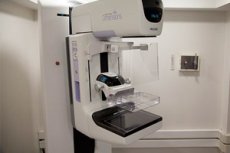
Mammography
Today, mammography is the main and most effective method of early diagnosis of breast cancer. The procedure is a screening examination and is carried out on special devices that allow identifying pathological growth and changes in tissues. The resulting images are compared with images of a healthy breast. During mammography, the breast is compressed with plates in order to take images from different angles. The organ tissue in the image is white, the fatty tissue is transparent, and the seals and pathological areas are clearly outlined.
Mammography makes it possible to recognize primary and secondary signs of a malignant process.
- Primary signs are microcalcifications and a tumor shadow that has a star-shaped or irregular shape with uneven contours. The tumor may be accompanied by a path to the nipple and cause its retraction, the skin is thickened and has ulcers. If there are microcalcifications on the breast, that is, calcium salt deposits in the walls of the gland ducts, then this indicates a high probability of a malignant process in the breast.
- Secondary signs are characterized by a variety of symptoms that manifest themselves in changes in the skin, nipples and tissues surrounding the neoplasm.
Computer tomography
CT of the mammary glands allows tracking the spread of tumor foci and metastases. It is performed both for early diagnosis of breast cancer and during the entire course of treatment, to monitor the results of therapy.
[ 10 ]
Magnetic resonance imaging
MRI of the mammary glands is performed using a powerful magnetic field. The mammary glands are irradiated with electromagnetic waves. As a result, the electromagnetic energy is recorded by special sensors and processed by a computer.
Positron emission tomography
It is a radionuclide tomographic method that quickly and accurately determines the presence of tumor processes. During the diagnostic process, a radiopharmaceutical containing radionuclides with positron beta decay is introduced into the glands.
PET allows us to establish the presence of a malignant neoplasm, detect metastases, determine whether cancer cells remain after treatment, and distinguish cancer from benign diseases and inflammatory processes.
Biopsy for breast cancer
A biopsy is a sample taken from a tumor with subsequent cytological examination. The advantages of this method are its low cost, ease of implementation and painlessness. Very often, a biopsy is performed under ultrasound monitoring. A biopsy can be performed by surgical intervention or by sectoral resection. The procedure is performed in an outpatient setting with anesthesia and does not require special preparation.
A diagnostic study allows identifying the type of neoplasm. A biopsy identifies hormone-dependent tumors using immunohistochemical tests. The treatment method and prognosis for recovery depend on the hormonal status of the tumor. A biopsy helps determine the histogenesis of a malignant neoplasm and develop an effective treatment plan, and determine the sensitivity of cancer cells to various types of therapy.
- Fine needle aspiration biopsy is the simplest and fastest diagnostic method. It is performed using a thin needle, so it does not require anesthesia.
- Trepan biopsy – performed in an outpatient setting, but with anesthesia. The needle is inserted into the tissue under ultrasound control.
- Surgical biopsy – used in cases where theran biopsy and fine-needle biopsy did not confirm the presence of oncology. The procedure is performed under full hospitalization and general anesthesia. Doctors perform surgery and examine the removed tumor.
Thermography
A diagnostic method that involves measuring the temperature of the skin of the chest. The study is based on the difference in temperature values between tumor and healthy tissue. Since the tumor has a large number of blood vessels, they emit heat, which can be detected using thermography.
This diagnostic method has not become widely used because it has a large number of false negative results.
Light scanning
Refers to the most modern diagnostic methods. The essence of the study is that infrared color is passed through the breast tissue, which allows determining the presence of tumor nodes and metastases.
The only disadvantage of this method is its increased sensitivity and insufficient specificity.
Galactophorography
It is performed in the presence of bloody discharge from the nipples. A contrast agent is injected into the milk ducts, which reveals multiple and single papillomas and intraductal cancer.
Pneumocystography
During this diagnostic examination, gas is introduced into the affected cavity of the mammary gland. The method makes it possible to identify intracystic pathological growths.
In addition to the above-described methods of cancer diagnostics, the patient undergoes an X-ray examination of the lungs, CT of the abdominal cavity and chest organs, ultrasound of the lymph nodes and abdominal organs. General clinical tests and examinations, as well as radioisotope studies of the skeleton, i.e. scintigraphy, are mandatory.
Such diagnostics will help to assess the extent of the spread of a malignant tumor in the body, the presence or absence of metastases in the lymph nodes and other organs. The results of the studies allow us to determine the characteristics and features of the pathology, as well as the state of the body. Please note that the breast cancer tumor marker CA15-3 is used to monitor the course of the disease, control treatment and detect relapses.
Breast Cancer Tests
Tests for breast cancer provide an opportunity to learn the characteristics of the pathological disease. Based on the results of the study, the doctor diagnoses one or another form of breast cancer and the stage of the tumor process. To conduct tests, blood and tissue from the affected area are taken from the patient.
- Timely diagnostics and testing allow us to identify and prevent recurrence of malignant neoplasms, and check the body for the presence of cancer cells after surgery or therapy.
- The tests allow us to detect a tumor at the earliest stages and determine whether a person is at risk.
When analyzing blood, tumor markers are studied: CA 15-3, CA 125 II, CYFRA 21-1, CA 72-4 and carcinoembryonic antigen (CEA). If these indicators are higher than normal, this may indicate both a malignant and a benign tumor.
Mucin-like cancer antigen CA 15-3 is found in the membranes of cancer cells. The norm is considered to be values from 0 to 26.9 U/ml. Blood tests are taken dynamically, this allows us to determine the rate of tumor growth, the risk of metastasis and relapse of malignant neoplasm. An additional confirmatory analysis is alpha-fetoprotein. The normal AFP value is considered to be from 0 to 7.51 U/ml. Deviations from the norm can indicate pathological processes in the body.
When examining tissues, an immunohistochemical analysis for breast cancer is performed. To perform it, special reagents are used that contain antibodies with special substances and breast tissues that are taken using a biopsy. The analysis is based on the antigen-antibody reaction. Thus, when foreign agents enter the body, a reaction occurs in the blood that blocks them. Immunohistochemical analysis allows you to identify the desired antigen of a cancerous tumor, so its implementation is very important for making a treatment plan.
Tumor markers for breast cancer
Tumor markers for breast cancer are proteins that circulate in the blood. The protein level increases in oncological diseases. But an increased concentration can also be observed in the body of a healthy person. That is why tumor markers are used as an auxiliary diagnostic method that allows you to detect relapses of the disease and evaluate the effectiveness of treatment.
For example, the tumor marker CA 15-3 is a marker of breast and ovarian cancer, and CA 125 is a marker indicating recurrence of tumor processes in the breast and ovaries. TRU-QUANT and CA 27-29 markers are also used for diagnostics.
An elevated level of this marker indicates a cancer process. Thus, when diagnosing carcinoma, the specificity level of CA 15-3 is 95%, unlike benign neoplasms. CA 15-3 is directly proportional to the size of the tumor lesion. That is, elevated values may indicate the involvement of lymph nodes in the oncological process. If the tumor marker increases by 25%, this indicates the progression of a malignant neoplasm. If the level decreases, the treatment is effective and the cancer recedes.
CA 15-3 allows you to find out about the presence of metastases and recurrence of the disease. If breast cancer was treated with chemotherapy or radiotherapy, the CA 15-3 values may temporarily increase. This indicates that the tumor is being destroyed, i.e. the therapy is effective. However, there have been cases where CA 15-3 had increased values in pregnant women, but this was not a sign of a malignant neoplasm.
- Tumor markers CA 15-3 and CEA
These tumor markers allow monitoring tumor development. Very often, CA 15-3 is performed simultaneously with a cancer-embryonic agent, i.e. CEA, which is a marker of rectal oncology.
The normal value of CA 15-3 is considered to be from 0-27 U/ml. If the tumor marker values are higher than the specified norm, this indicates a metastatic process. The norm of CEA is from 0-5 U/ml. Decoding of tumor markers should be carried out only by a doctor. Cancer is not diagnosed only by an elevated level of CA 15-3 or another tumor marker. To confirm the disease, a comprehensive diagnosis is carried out.
Breast cancer diagnostics involves a comprehensive approach to identifying a malignant neoplasm, determining its type and other features of the disease. The choice of treatment methods depends on the timeliness of diagnostics and its effectiveness. The earlier cancer is detected, the better the prognosis and the higher the chances of recovery.