Medical expert of the article

New publications
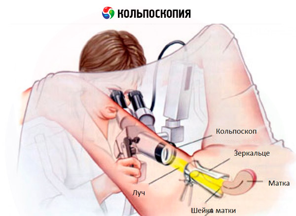
Colposcopy
Last reviewed: 23.04.2024

All iLive content is medically reviewed or fact checked to ensure as much factual accuracy as possible.
We have strict sourcing guidelines and only link to reputable media sites, academic research institutions and, whenever possible, medically peer reviewed studies. Note that the numbers in parentheses ([1], [2], etc.) are clickable links to these studies.
If you feel that any of our content is inaccurate, out-of-date, or otherwise questionable, please select it and press Ctrl + Enter.
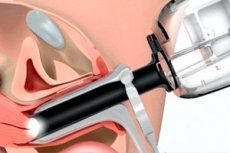
The colposcopy was proposed in 1925 by Hinzelman. Colposcopy allows you to make a detailed examination of the vaginal part of the cervix and vaginal walls with a special optical device - the colposcope. The design of the colposcope includes an optical system of lenses with a focal length of 25-28 cm and interchangeable eyepieces, giving an increase from 6 to 28 times. Modern colposcopes have a photographic device that allows you to document research data.
Some models of the colposcope make it possible to carry out studies using the fluorescence analysis method - by detecting secondary luminescence in ultraviolet rays.
Indications for conduction
Diagnostic and differential diagnosis of the pathological changes in the vaginal part of the cervix, the walls of the vagina and the vulva.
In the gynecological practice, the following types of colposcopic examination are consistently performed.
Types of colposcopy
Simple colposcopy - examination of the cervix, which is indicative. Determine the shape, size of the neck and outer throat, color, mucosal relief, the border of the flat epithelium covering the cervix, and the cylindrical epithelium of the cervical canal.
Extended colposcopy - examination after neck treatment with 3% acetic acid solution, which causes a short-term epithelial edema, swelling of the cells of the subulate layer, reduction of subepithelial vessels and a decrease in blood supply. The action of acetic acid lasts 4 minutes.
After studying the colposcopic pattern of the neck, treated with acetic acid, a so-called Schiller test is performed-neck lubrication with a cotton swab dipped in 3% Lugol's solution. The iodine contained in the solution stains glycogen in the cells of the healthy, unaffected flat epithelium of the neck in a dark brown color. Thin cells (atrophic age-related changes), as well as pathologically altered cells with epithelial dysplasia, are poor in glycogen and iodine solution are not stained. Thus, the zones of the pathologically altered epithelium are identified and the areas for biopsy are planned.
Colposcopic microscopy. In vivo histological examination of the vaginal part of the cervix. It is produced by a contrast luminescent colpomicroscope, the tube of which is brought directly to the cervix; increase up to 300 times. Before inspection, the cervix is stained with a 0.1% solution of hematoxylin. In the colpomicroscopy of the unchanged cervix of the flat epithelial cells covering it, they have a polygonal shape, with clear boundaries, the cell nuclei are colored violet, the cytoplasm in blue; Podopithelialnye vessels, visible at a depth of 70 microns, have a straight line and uniform division, the bed of them is not widened. The colpomicroscopic method of investigation has a high accuracy of revealing pathological changes, the coincidence of this method with the results of histological examination of the cervix is 97.5%.
Chromocolposcopy is a modification of an advanced colposcopy in which the cervix is stained with various dyes (methyl violet, 0.1% hematoxylin solution, 1% solution of toloidin blue). The difference in the color of the flat and cylindrical epithelium allows us to clarify the pathological process and its outer boundaries.
A variety of advanced colposcopy is the study of the colposcopic picture of the vaginal mucosa of the cervix through green and yellow filters, as well as examination in ultraviolet rays to reveal more precise contours of blood vessels.
Fluorescence colposcopy - examination of the cervix in ultraviolet rays after staining it with fluorochrome (intravital method of histochemical examination of tissues using ultraviolet rays). As a fluorochrome used uranium in the 1:30 000 dilution. Normal mucosa is characterized by a dark blue and purple glow. In the early forms of cancer, there is a bright yellow, salad-yellow, crimson glow. With severe cancer with necrosis and hemorrhages complete extinguishing of fluorescence is observed. The coincidence of diagnoses in fluorescent colposcopy with histological data is noted in 98% of cases.
Colposomyroscopy is the most perfect method of examining the vaginal part of the cervix, allowing it to be viewed with an increase of 175-280 times. This is an in vivo histological study of the cervical tissue in the incident light. When studying the epithelial cover and features of cellular structures, the cervix is stained with 0.1% aqueous solution of hematoxylin. Usually, a targeted colpomicroscopy is used, which is based on the staining of suspicious areas revealed during colposcopy.
The advantage of colpomicroscopy is that it is a completely harmless and painless method that allows studying the morphological changes in the surface of the cervix in dynamics both in norm and in pathology. This method is highly reliable.
The disadvantage of the method is that it allows us to judge only the state of the surface layers of the epithelium and does not allow detection and differential diagnosis of intraepithelial carcinoma and invasive cancer. The method is not sufficiently informative in case of lesion of the cervical canal. It can not be used for narrowing the vagina, bleeding tissues, necrotic changes in the cervix.
Luminescent colpomicroscopy is an advanced method of colposcopy, supplementing the survey data and expanding the possibilities of topical diagnosis.
Explanation of results
The colposcopic method of cervical examination has high accuracy in the detection of precancerous and cancerous diseases of the cervix, in the diagnosis of cervical endometriosis, polyps, endocervicitis.
The normal epithelium in colposcopy appears smooth, shiny, light pink in color, and after treatment with Lugol's solution, the neck acquires a uniform brown color.
Benign colposcopic changes include ectopia, a transformation zone, true erosion, changes associated with colpitis and diathermo-coagulation, which was earlier transferred.
Atypical colposcopic pictures include leukoplakia, the basis of leukoplakia, the papillary base, fields, a typical transformation zone and atypical vessels.
Ectopia is characterized by a pattern with the formation of papillae with loop-shaped vessels in them. The transformation zone is the area of the cervix, on which the prismatic epithelium is replaced by a multilayered flat epithelium. In appearance, these are smooth areas near the papillae of ectopy, against which the mouths of the glands are located. True erosion is a site of the vaginal part of the cervix, devoid of epithelial cover. When colpitis on the walls of the neck and vagina, many small blood vessels are seen.
Leukoplakia - shiny white spots, sharply delimited from the surrounding mucous membrane, iodnegative when treated with Lugol's solution.
The basis of leukoplakia - grains of red color on a white or yellowish background, iodnegative. Fields - white or yellowish polygonal areas, separated by thin red borders, iodnegative.
Atypical zone of transformation are different combinations of atypical epithelium, also iodnegative. Atypical vessels are arranged randomly, have a bizarre shape, there are no anastomoses between them. When the Schiller assay does not disappear, as with benign changes, but become more clearly visible.
For precancerous conditions characterized by the presence of atypical epithelial, located at different width, strong keratinization and atypical condition mucosa.
With preinvasive cancer, atypism of blood vessels is noted, with microcarcinoma - a chaotic arrangement of blood vessels, inhomogeneity of the relief.
 [
[