Medical expert of the article

New publications
Atresia of the esophagus
Last reviewed: 23.04.2024

All iLive content is medically reviewed or fact checked to ensure as much factual accuracy as possible.
We have strict sourcing guidelines and only link to reputable media sites, academic research institutions and, whenever possible, medically peer reviewed studies. Note that the numbers in parentheses ([1], [2], etc.) are clickable links to these studies.
If you feel that any of our content is inaccurate, out-of-date, or otherwise questionable, please select it and press Ctrl + Enter.
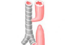
Atresia of the esophagus is a congenital malformation in which the esophagus blindly ends at a distance of approximately 8-12 cm from the entrance to the oral cavity.
Atresia of the esophagus (Q39.0, Q39.1) is the most common malformation in the neonatal period and diagnosed immediately after birth. The malformations listed below manifest later, often complicated by aspiration pneumonia, hypotrophy, esophagitis.
Atresia of the esophagus is an incomplete formation of the esophagus, often combined with the tracheoesophageal fistula. Diagnosis is made when it is impossible to carry out a nasogastric tube into the stomach. Treatment is prompt.
Congenital tracheal-esophageal fistula without atresia is a pathological canal lined with granulation tissue or epithelium, reporting unchanged lumen of the esophagus with the lumen of the trachea.
Atresia of the esophagus is the most common variant of atresia of the gastrointestinal tract. There are 5 main types of esophageal atresia. The most common type (85%) is when the upper esophagus ends blindly, and the tracheoesophageal fistula opens to the lower. The next most common (8%) type is pure esophageal atresia without fistula formation. The remaining options include fistula "H type" between the trachea and the esophagus, the esophagus pass (4%), esophageal atresia with proximal tracheoesophageal fistula (1%) and esophageal atresia with two fistula (1%).
ICD-10 code
- Q39 0 Atresia of the esophagus without fistula.
- Q39 1 Atresia of the esophagus with tracheoesophageal fistula.
- Q39.2. Congenital tracheal esophageal fistula without atresia.

Epidemiology
Epidemiology of esophagus atresia
The frequency is 1 per 3000-5000 newborns. About 100 variants of this defect are known, but there are three most common ones:
- esophagus atresia and fistula between the distal part of the esophagus and the trachea (86-90%),
- isolated esophageal atresia without fistula (4-8%),
- tracheoesophageal fistula, "type H" (4%).
In 50-70% of cases of esophageal atresia, there are concomitant malformations:
- congenital heart defects (20-37%),
- gastrointestinal defects (20-21%),
- defects of the genitourinary system (10%)
- defects of the musculoskeletal system (30%),
- defects of the craniofacial region (4%).
In 5-7% of cases, atresia of the esophagus is accompanied by chromosomal abnormalities (trisomy 18, 13, and 21). A peculiar combination of developmental abnormalities in esophageal atresia is indicated as “VATER” after the initial Latin letters of the following malformations (5-10%):
- spinal defects (V),
- malformations of the anus (A),
- tracheoesophageal fistula (T),
- esophagus atresia (E),
- defects of the radius (R).
30-40% of children with esophageal atresia are not full term or have intrauterine growth retardation.
Atresia of the esophagus is a very rare pathology, diagnosed in 4% of children with atresia of the esophagus.
Causes of the esophagus atresia
Causes of esophageal atresia
Atresia of the esophagus occurs when there is a mismatch between the direction and rate of growth of the trachea and esophagus, as well as the processes of vacuolization of the esophagus in the period from the 20th to the 40th day of intrauterine development.
Symptoms of the esophagus atresia
Symptoms of esophageal atresia
In the first hours of life, there is abundant discharge of foamy saliva from the mouth and nose of the child, sometimes vomiting. Symptoms of respiratory failure (shortness of breath, wheezing in the lungs, cyanosis) appear as a result of the aspiration of mucus from the oral end of the esophagus into the trachea and the discharge of gastric contents into the respiratory tract through the lower tracheoesophageal fistula. In some children with distal tracheoesophageal fistula, abdominal distension is observed due to the release of air into the stomach, especially after the use of mechanical ventilation. In the absence of a distal tracheoesophageal fistula in a child, a sunken belly is noted.
Characteristic signs are regurgitation, coughing and cyanosis after feeding attempts and aspiration pneumonia aspiration pneumonia. Atresia of the esophagus with a distal fistula leads to an increase in the abdomen, as when screaming, the air is pushed out of the trachea and through the fistula enters the lower esophagus and stomach. When probe feeding a child, all of the above symptoms disappear.
Where does it hurt?
What's bothering you?
Diagnostics of the esophagus atresia
Diagnosis of esophageal atresia
The diagnosis involves the inability to hold a nasogastric tube into the stomach. A radiopaque catheter determines the localization of atresia during radiography. In atypical cases, a small amount of water-soluble contrast may be required to determine the anatomy of a defect during fluoroscopy. Contrast material should be quickly evacuated, since its entry into the lungs can cause chemical pneumonitis. This procedure should be performed by an experienced radiologist at the center where the newborn will be operated.
In atresia of the esophagus, it is impossible to introduce a probe into the stomach. The probe encounters an obstacle at a distance of about 10 cm from the bite line. Given the immense importance of early diagnosis, the sensing of the esophagus should be carried out to all babies who have frothy discharge from the mouth and respiratory disorders after birth. On the radiograph of the chest and abdominal cavity with the introduction of the radiopaque probe until it stops in the esophagus, a blindly ending upper segment is detected. Detection of air accumulation in the stomach and intestines is pathognomonic for the tracheoesophageal fistula in the lower esophagus. Lack of air is characteristic of vaginal atresia of the esophagus. Contrasting of the esophagus increases the frequency of respiratory complications and deaths.
Prenatal diagnosis
Prenatal diagnosis is based on indirect signs, such as:
- the high water flow associated with a decrease in the circulation of amniotic fluid due to the inability of the fetus to swallow amniotic fluid,
- the absence of an echographic image of the stomach or the small size of the stomach during dynamic ultrasound observation.
The sensitivity of these signs is 40-50%. In the II – III trimester, it is possible to visualize the periodically filling and emptying blind proximal end of the esophagus with an accuracy of 11–40%.
What do need to examine?
Who to contact?
Treatment of the esophagus atresia
Treatment of esophageal atresia
Preoperative preparation
If an esophageal atresia is detected, it is urgent to transfer the child from the maternity hospital to the surgical hospital.
In order to reduce the aspiration syndrome, it is necessary to give the child an elevated position (30-40 °), insert a catheter attached to the system of constant aspiration into the proximal end of the esophagus, or carry out frequent aspiration of mucus from the oropharynx.
It is necessary to cancel feeding and start infusion therapy, prescribe broad-spectrum antibiotics, hemostatic drugs. Inhalation of humidified oxygen helps to eliminate hypoxia. With an increase in respiratory failure, tracheal intubation is performed and mechanical ventilation is started. With traditional mechanical ventilation in a child with a distal tracheoesophageal fistula, a significant discharge of air through the fistula into the stomach is possible, which leads to over-inflation of the stomach and intestinal loops. Enlarged abdominal organs limit the excursion of the diaphragm, as a result of respiratory failure progresses, possible perforation of the stomach and cardiac arrest. In these cases, you should try to change the position of the endotracheal tube to rotate or deepen to reduce air discharge through the fistula. If ventilation is not possible due to a significant discharge of air through the fistula, an AChO IVL or single lung ventilation is indicated. With a significant air discharge, the issue of emergency surgery of gastrostomy and / or ligation of the tracheoesophageal fistula should be resolved.
If the child’s body weight is more than 2 kg, and there are no violations of the vital systems of the body, the operation begins immediately after the necessary research has been carried out. If the child’s body weight is less than 2 kg, or there are disturbances of homeostasis and other malformations, the duration of the preoperative preparation is increased and correction of the identified violations is carried out.
At the same time, they conduct diagnostic activities aimed at identifying concomitant malformations and disorders of the vital organs:
- determination of blood group and Rh factor
- CBS definition
- clinical blood test,
- general urine analysis,
- blood chemistry,
- coagulogram analysis
- EcoG,
- EKG,
- Ultrasound of internal organs
- neurosonography.
Venous access is carried out by catheterization of the central vein, while it is preferable to hold the catheter into the superior vena cava from the ulnar or axillary vein. It is not recommended to perform puncture and catheterization of the right right subclavian and internal jugular veins because of the high risk of hematoma formation in the area of operation.
Preoperative treatment of esophageal atresia is aimed at ensuring that the child is in optimal condition before the operation, as well as prevention of aspiration pneumonia, which makes the operation more dangerous. Refrain from oral feeding. Constant suction through a double lumen catheter in the upper esophagus prevents aspiration of swallowed saliva. child should be in a prone position with the head raised 30-40 ° and the right side of the body lowered to facilitate emptying the stomach and minimize the risk of aspiration of the acidic contents of the stomach through the fistula. If radical surgery is necessary to postpone due to deep prematurity, aspiration pneumonia, or other congenital malformations, a gastrostoma should be formed for decompression of the stomach. Suction of gastric contents through the gastrostomy tube reduces the risk that it enters the tracheobronchial tree through the fistula.
Surgical treatment of esophageal atresia
Surgery for esophageal atresia refers to urgent surgical interventions.
In more than 90% of cases with esophageal atresia, a fistula is isolated and ligated with an overlay of esophagoesophagoanastomosis. During the operation, a nasogastric tube is passed through the anastomosis, which must be carefully fixed. In the mediastinum leave drainage attached to the passive aspiration system.
In case of a lumpless form due to a large diastasis between the atreated ends of the esophagus, it is often imposed on the gastrostomy and esophagostomy. In atresia of the esophagus, thoracoscopic surgery is possible.
When the child's condition stabilizes, an extrapleural surgical correction of esophageal atresia and closure of the tracheoesophageal fistula can be performed. Sometimes the atresia of the esophagus requires the esophagoplasty to be performed by the colon section.
Postoperative period
Management tactics depend on the severity of pulmonary disorders, concomitant anomalies, and degree of prematurity.
Extubation immediately after surgery is possible in full-term infants without concomitant developmental anomalies and severe lung damage.
When there is a risk of respiratory failure after surgery, the child is given a mechanical ventilation. Ventilation support is stopped as soon as the child is able to independently provide gas exchange and respiration.
Tracheomalacia is a common anomaly in tracheoesophageal fistula. It can cause cyanosis and apnea after extubation of the child. In such cases, tracheostomy or tracheopexy is indicated.
In the next 3-7 days after the operation, the neck should not be unbendable, as this will stretch the esophageal anastomosis and there may be a failure of the stitches.
During the aspiration of mucus from the trachea, the catheter is inserted strictly to the depth of the endotracheal tube, in order to avoid recanalization of the fistula. It is necessary to constantly carry out the removal of nasopharyngeal contents without inserting a catheter into the esophagus beyond the anastomosis.
Drainage, left in the mediastinum, is removed on the 5-7th day, if during this time there is no pathological discharge. In some cases, before the drainage is removed, an x-ray examination of the esophagus with a water-soluble contrast is performed.
Anesthesia after surgery is carried out by infusion of opioid analgesics [fentanyl at a dose of 2-5 μg / (kgkh), trimeperidine at a dose of 0.05-0.2 mg / (kgkh)] in combination with metamizole sodium (at a dose of 10 mg / kg) or paracetamol (10 mg / kg) for 3-5 days, then they are transferred to bolus administration of these drugs according to indications.
Infusion therapy in the postoperative period is carried out at the rate of physiological needs. On the first day after surgery, 5-10% glucose solution is used, to which electrolytes are added. After 12-24 h after the operation with uncomplicated postoperative period, parenteral feeding begins.
Antibiotic therapy in the postoperative period is carried out, focusing on monitoring the microecological status of the child, but in the coming days after the operation, metronidazole IV is shown at a dose of 15 mg / (kg-day).
[30], [31], [32], [33], [34], [35], [36], [37], [38]
Feeding a child with esophageal atresia
If a gastrostomy is imposed and the feeding catheter is placed in the duodenum, feeding can begin 24 hours after surgery.
When imposing direct esophagoesophagous anastomosis, nutrition is carried out through a nasogastric tube starting from the 5-7th postoperative day.
Oral feeding is possible only 7–10 days after surgery.
With esophageal atresia, enteral nutrition begins with anti-reflux mixtures (Frisom, Nutrizon anti-reflux, Humana AR) in combination with prokinetics (domperidone at a dose of 0.5 ml / kg), since this developmental defect after the operation usually results in gastro-esophageal reflux.
Complications of esophageal atresia
The most frequent acute complications are the failure of the anastomosis and the formation of strictures. After successful surgical correction, feeding difficulties are often encountered due to impaired motility of the distal esophagus, which predisposes to the development of gastroesophageal reflux (GER). If drug treatment for GER is ineffective, Nissen fundoplication may be required.
Complications in the immediate postoperative period:
- pneumonia,
- failure of the anastomosis,
- mediastenit,
- laryngotraemalization,
- gastroesophageal reflux,
- anemia.
Forecast
Esophageal Atresia Prediction
Survival with isolated atresia of the esophagus is 90-100%, with severe combined anomalies - 30-50%. In uncomplicated forms of esophageal atresia, the prognosis is favorable. In the coming years after surgery, dysphagia and eating disorders associated with gastroesophageal reflux or the development of esophageal stenosis may occur. Increased risk of respiratory infections, pneumonia, asthma due to microaspirations of gastric contents into the trachea.