Medical expert of the article

New publications
Angioma
Last reviewed: 17.10.2021

All iLive content is medically reviewed or fact checked to ensure as much factual accuracy as possible.
We have strict sourcing guidelines and only link to reputable media sites, academic research institutions and, whenever possible, medically peer reviewed studies. Note that the numbers in parentheses ([1], [2], etc.) are clickable links to these studies.
If you feel that any of our content is inaccurate, out-of-date, or otherwise questionable, please select it and press Ctrl + Enter.
What is meant by the term "angioma"? This type of benign tumor, consisting of blood vessels or lymphatic drainage.
Pathology occurs as a consequence of the expansion and modification of the vascular walls. The severity of the disease depends on the location of the angioma, as well as on its size and the degree of influence on the flow of fluid inside the vessels.
Causes of the angiomas
Angioma develops as a result of proliferation of the vasculature and endothelial tissue. A typical sign of such formation is formation of fine-vascular bonds in the arteriolar and venule transitions. As a result, there arises a peculiar shunting of blood flow past the capillary network, which explains the appearance of the morphological and clinical picture of angioma.
The disease can develop for various reasons, but most often it is a congenital anomaly. Even if the child does not show such a pathology, then she can express herself in adulthood.
Is angioma dangerous?
Angiomas are any benign tumors of vascular origin, regardless of which system they represent - the circulatory or lymphatic. Neoplasms can be located in the superficial layer of cutaneous or mucous membranes, in muscle tissue, in cavities and tissues of internal organs, in the brain. Depending on where this tumor is located, and the degree of its danger depends. Significant angiomas in size, which are formed in vital organs, really represent a danger, and above all, the probability of bleeding.
Vascular formations that appear on the surface of the skin are very similar to moles: their danger is not so great.
The risk of bleeding may also depend on the type of education. We will talk about such varieties below.
Risk factors
The theory of congenital vascular anomalies is still the guesswork of scientists. Reliable risk factors for the development of angiomas have not yet been determined. Among other possible causes, too, are insolation, women's diseases during the period of childbearing, hormonal imbalance, liver disease, etc.
Symptoms of the angiomas
The clinical symptoms of angiomas may differ, depending on the type of neoplasm, its location, volume and developmental features.
In most cases, the first signs are found even during the baby's newborn period, or during the first year of his life. According to statistics, in girls the disease manifests itself more often. Only a few months after the appearance of tumors can increase from a few millimeters to 2-3 centimeters. In addition, the number of pathological elements that can arise anywhere: on the surface of the skin, in the oral cavity, in the groin, in the respiratory and musculoskeletal system, the liver, etc., can also increase. Depending on the location, the presence of vascular neoplasms can provoke frustration digestion, respiration, urination, defecation, etc.
Angioma of the bone can appear in the vertebrae, on the tubular bones and bones of the cranium, which is often accompanied by pain syndrome, deformity, periosteum damage.
Vascular brain tumors are considered extremely dangerous: they can provoke disorders of brain functions and hemorrhages in the membranes of the brain.
Angiomas from lymphatic vessels are most often formed on the skin. In most cases, they are found in the neck, in the mouth, on the face, under the arms, on the genitals. Such tumors have the appearance of a painful compaction, which can reach considerable sizes. Lymphangioma grows relatively slowly over several years.
Spinal Cord Angioma
The clinical picture of angioma of the spinal cord may differ, depending on where the tumor lies on which part of the spinal column. The location is distinguished by:
- vascular tumors with craniospinal localization (from the cranium to the spinal cord);
- angiomy of the cervical department;
- angiomy of the thoracic region;
- lumbosacral angiomas;
- vascular tumors of the sacrococcygeal zone.
Symptoms develop as a result of pressure on the nerve endings and the spinal cord. Among the main signs more often appear:
- pain (with irradiation, constant or intermittent);
- increased sensitivity of the skin in the zone of innervation of the nerve;
- paresthesia;
- weakening of the muscles of the back;
- disorganization of the work of some internal organs.
The listed signs are characteristic not only for angiomas, but also for some other tumoral processes in the spinal column. Therefore, a diagnosis can be made only after a thorough diagnosis.
Angioma of the frontal lobe
The frontal lobe of the brain is approximately 29% of the entire cortex, and its weight is more than half the total mass of the brain. The frontal lobe is responsible for movements, for speech skills, for individuality and decision-making. Accordingly, when the angioma reaches a large size, painful symptoms may appear:
- dizziness;
- headache;
- impaired motor coordination;
- lack of interest in what is happening;
- speech disorders, inhibition, lethargy.
At the initial stages of development, symptomatology is weak. Sometimes signs of the disease occur only when the tumor starts bleeding.
As a result of excessive accumulation of vessels, their splicing and thinning of the walls of the droppings, a hemorrhage appears, which is symptomatic of a stroke. There are convulsions, paralysis, paresis, vision impairment, speech disorders, etc.
 [27], [28], [29], [30], [31], [32], [33], [34]
Angioma of the cerebellum
Symptoms of angiomy of the cerebellum can be the same as in the lesion of the frontal lobe. In addition, the disease can be hidden if the tumor is small and does not press on the surrounding tissue. Problems can be detected only after the angioma begins to bleed.
Progression of the vascular tumor is accompanied by a circulatory disorder in the affected area, which is explained by the accumulation and interweaving of blood vessels of different calibers. The outcome of the disease largely depends on the overall state of the vascular system in the body. For example, with hypertension and increased blood pressure, the risk of hemorrhage rises several times.
Especially dangerous are multiple hemorrhages, which can lead to irreparable negative consequences.
[35], [36], [37], [38], [39], [40], [41], [42], [43]
Angioma of the eye
Vascular changes can affect not only the skin surface of the body and the brain, but also organs, including the eyes.
Angioma of the retina is mainly considered an innate disease. The plexus of the vessels is detected at birth, or somewhat later. Often the problem is determined when the person gradually begins to deteriorate vision, up to complete blindness.
Angioma of the retina occurs with the formation of weak vascular interlacing of a different color shade - from crimson to grayish-greenish. Around the tumor, sometimes there is a puffiness center and small areas of bleeding.
Vascular neoplasms of the eyes are characterized by slow development with normal preserved visual function. Most often, only one eye is affected.
Further progression of the disease leads to the appearance of cataracts or retinal detachment.
Throat Angiomy
The vascular tumor in the pharynx resembles a different size of a burgundy or brownish nodule on a broad base. The standard location of the angioma is the area of the soft palate and the palatine arch, the root of the tongue, the pharyngeal walls and tonsils.
Neoplasm can grow to a considerable extent. The first signs are manifested by the diffuse sensation of a foreign body in the pharynx and bleeding, although in some cases there may not be symptoms. Most often bleeding occurs after eating rough food, which traumatizes the vascular bundle. Damage to a large size can be accompanied by serious bleeding, up to a lethal outcome.
Common symptoms can be divided according to the affected pharyngeal department:
- when the upper division is affected, there may be problems with ingestion of food, perspiration and coughing;
- with the defeat of the middle section, hoarseness in the voice, blood veins in the salivary fluid is detected;
- with the defeat of the lower part, difficulties arise with breathing in air and speech.
There were no exact causes of angiomy of the pharynx. Specialists assume a hereditary etiology of the disease.
Angiomas for HIV
Patients with the immunodeficiency virus often develop diseases of the cardiovascular system. Vascular lesions can be suspected in patients with blood pressure changes, with frequent inflammatory processes in the joints and muscles, with diseases of the urinary system and the nervous system, with myocardial and cerebral ischemia.
Angiomas with HIV are not detected more often than in other people. In some cases, vascular neoplasms are formed as a result of an inflammatory reaction in the walls of the vessels, mainly in patients from 20 to 30 years, regardless of the presence of atherosclerotic changes. In this case the peripheral network of capillaries is more often affected.
The symptomatology of angioma does not differ from that of the rest of the patients. The disease is detected during angiography, or in the presence of characteristic signs of pathology.
Patients with angiomas on the background of HIV should be regularly examined by a specialist, since the risk of bleeding in such patients is much higher.
Angioma in newborns
Angioma in most cases has an innate etiology. There is a connection between some factors that arise during pregnancy and the development of vascular tumors. Thus, neonatal angiomas can form as a result of the following reasons:
- violations of intrauterine development of the fetus during the vascular network (this occurs already in the third week of pregnancy);
- infectious diseases of a woman in the period of gestation;
- risk of spontaneous abortion.
In the presence of angiomas in a newborn child, oncologist's advice and supervision are considered mandatory. If you ignore the signs of angioma, you can miss unpleasant complications in the form of bleeding. There is also a certain risk of degeneration of the vascular tumor into a malignant formation with aggressive course. Therefore, most often in early childhood practice the removal of suspicious vascular congestion.
[49], [50], [51], [52], [53], [54], [55], [56], [57], [58]
Angioma in Pregnancy
As you know, during the period of bearing the baby in the female body, a huge number of changes occur, which is mainly due to the redistribution of the level of hormones. At this time, often there is excessive pigmentation of the skin, and the appearance of angiomas is not uncommon.
Vascular formations can be found on the face, in the decollete zone, on the forearms. The capillary network in pregnant women becomes particularly vulnerable: hence the appearance of vascular asterisks and stellate hemangiomas.
Some such formations can disappear on their own in the postpartum period, when the hormonal background of the mother comes back to normal. However, you should closely monitor the tumor: even a slight change in the shade or size of the angioma should alert and cause a medical consultation.
It is also necessary to avoid possible injury to the neoplasm. Even a small vascular overgrowth can provoke severe bleeding.
[59], [60], [61], [62], [63], [64], [65], [66], [67], [68], [69]
Forms
Angiomas are primarily divided into those that develop in the circulatory system (hemangioma), or in the lymphatic system (lymphangioma).
Classification by histological features:
- monomorphic angioma - formation on the basis of any one vascular element;
- polymorphic angioma - formation from several vascular elements.
Classification by structural feature:
- Capillary angioma is the most common type of disease in which capillaries are the basis of the structure. Capillary formation is often located on the surface of the skin, less often - in the organs inside the body.
In most cases, capillary formation is detected during the baby's neonatal period. Angioma has the ability to expand and increase in size, but by the time the body grows, the angiomy gradually fades and disappears.
Self-elimination of the tumor occurs as follows:
- the smallest vessels entering the structure of education, stick together and cease to flow blood;
- the tumor is discolored, its size decreases;
- there is destruction of capillaries;
- the tumor is not visually determined.
It is worth noting that the further development of capillary angioma is unpredictable. Sometimes it is able to grow and expand to the nearest vessels.
- Venous angioma is detected much less frequently, in contrast to capillary angioma. As it becomes clear from the title, such a tumor consists of a venous vascular network, which, growing, acquires a bluish tinge. Venous angioma can be quite large. It affects both superficial and deep venous vessels.
- Cavernous angioma is an even more rare type of vascular neoplasm. Such a tumor is built on the basis of vessels with thin walls, in which specific areas of expansion - cavities are formed, where there may be a thrombus formation. There are cavernous tumors in the skin and digestive organs. Visually angioma of this type resembles an elevation of a bluish-red hue, which has a spongy structure. The caverns of the sponge are filled with blood fluid.
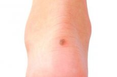
- Cherry angioma is a dermatological disease that affects people in adulthood (mostly after 30 years). The disease is characterized by the appearance of small reddish papules (1-5 mm) on the surface of the body, mainly in the chest area, or on the scalp. Such an angioma is a cluster of dilated blood vessels in the dermis layer.
Why cherry angioma is formed, is still unknown. Such formations are benign, but they tend to spread over the body with age. Multiple angioma is already a serious enough reason for contacting an oncologist.
- Stellate angioma (the second name is spidery angioma) can occur at any age: the disease has the form of a red formation, from which the threads of the same color - blood-filled capillaries branch off. Externally, stellate angioma does resemble an asterisk, or spider. The main vessel supplies nutrition to all affected tumor structures. Due to this, the formation gradually increases to 8-10 centimeters, only in rare cases without changing its size.
The most frequent localization of asterisks is the skin of the face, head and shoulders. The reason for this is seen in a sharp increase in the level of estrogen in the bloodstream, or in a genetic predisposition. Such tumors look unaesthetic, but do not cause their owner any unpleasant sensations.
- Spot angioma is the so-called "ordinary" angioma, which poses absolutely no danger to human health. The tumor has the appearance of a slight dense formation protruding above the surface of the skin. Color shade - from slightly pink to dark red or burgundy. In some cases, the dot elements do not add visual appeal, but experts do not advise them to be hastily removed - this can lead to further spread of the process.
- Glomusnaya tumor (glomus angioma) consists of arteries and veins, can be located independently or multitudinously. Glomus is a glomerular arteriovenous anastomosis (vascular connection). The vessels of such a tumor have a narrowed lumen and a large number of glomerular cells, which are regarded as altered structures of smooth muscle tissues.
The glomus of the angioma is located mainly on the toes and hands, sometimes on the limbs. They do not cause pain, although they develop close to the surface of the skin. In most cases, the disease occurs in children, and more often in boys.
- Senile angioma has the second name - senile. Hence it is clear that this type of disease is most typical for elderly patients. Often, the disease is mistaken for an ordinary birthmark, although its structure is somewhat different. Senile angiomas are not dangerous, even if they spread on the surface of the skin in large quantities. Usually they are considered as a kind of cherry angioma.
The form of the senile tumor is spherical, oval, or in the form of a hemisphere. The bulge has an uneven surface, ranging in diameter from 1 to 6 mm. It occurs mainly in fair-skinned people, regardless of gender.
- Flat angioma is a congenital disease. It looks like a speck of different shapes, a dense pink or purple hue. With physical exertion or temperature drop, the color depth of the tumor can vary.
Flat angioma is usually located in the face, neck, back, or upper limbs. This kind of neoplasms can behave unpredictably: sometimes tumors can pass into the active stage of growth and occupy quite extensive skin surfaces. In rare cases, tissues can be susceptible to necrosis, bleed, ulcerated. If this happens, then the consultation of the oncologist-dermatologist is mandatory and urgent.
Vascular angiomas are also subdivided depending on their location in the tissues of the body. Thus, distinguish angioma of the brain, skin, internal organs, etc.
For example, skin angioma is located, as a rule, in the surface layers. It can be seen with the naked eye. Such tumors usually do not touch, unless unless they give trouble to their owner. Skin angiomas are not dangerous. However, they must be protected from injuries and damage.
Unlike a superficial tumor, brain angioma is considered a more serious pathology and can cause the emergence of unpleasant symptoms such as headaches, seizures, nausea, etc. Moreover, the long-term progressive angioma of the brain can provoke hemorrhage and other negative consequences.
Complications and consequences
Of the most dangerous consequences of vascular tumors, bleeding is necessary first. The risk of hemorrhage is estimated at about 3% of annual cases, from minor bleeding to massive bleeding. Especially dangerous if such complications occur in the brain or spinal cord.
Prevent hemorrhage in advance is almost impossible. The vascular cluster may have a stable course, but sometimes the deterioration occurs suddenly. Similarly, it is not excluded and the sharp regression of the tumor, until its disappearance.
However, there are a number of factors that increase the risk of bleeding:
- tumor size;
- disturbance of blood flow in altered vessels;
- increased intravascular pressure;
- increase in the volume of circulating blood;
- already existing hemorrhages.
It was noted that the superficial location of the neoplasm is characterized by a lower ability to bleed. However, such hemangiomas require regular monitoring by a doctor.
Re-emergence, or relapse of angioma, may occur after a conservative, non-radical removal of the lesion. Virtually all vascular pathological clusters are prone to such a repeated development. Therefore, if angiomy delivers discomfort, then it is recommended to remove it radically, surgically.
Diagnostics of the angiomas
Surface cutaneous angioma is diagnosed directly with normal external examination and palpation. In this case, a characteristic feature is the blanching of the formation after pressing on its central region.
With a deep disposition of the pathology, a comprehensive diagnosis is prescribed. Among many diagnostic techniques, the following can be used:
- radiography of the bone system;
- angiography of the cerebral vasculature;
- angiography of other organs;
- lymphangiography;
- Ultrasound of blood vessels;
- consultation of the specialized specialist (otolaryngologist, urologist, neurologist, pulmonologist, etc.).
Standard blood tests are poorly informative. In the diagnosis of lymphangioma, puncture can be performed, followed by analysis of the intraluminal fluid.
What do need to examine?
How to examine?
Differential diagnosis
Differential diagnosis is performed with the following diseases:
- angiokeratoma;
- pigment nevus;
- melanoma;
- angioleiomioma;
- hemangiopericytoma.
Lymphangioma should be distinguished from isolated scleroderma and pachydermia.
Instrumental diagnosis is most often represented by angiography. This is the radiopaque method of investigation, which makes it possible to visualize the vascular network, its direction, size and other characteristics on the film. Recently, a more advanced method is often used - super selective angiography. This method differs from the previous one in that the contrast is not introduced into the common vascular bed, but directly into the vascular cluster.
Who to contact?
Treatment of the angiomas
The choice of method of treatment can depend on the type of angioma, on its location, size and flow characteristics. If the tumor is insignificant and does not cause the patient any discomfort, then it is most often not touched. With disturbing neoplasms, the most common methods are:
- removal by laser;
- electrocoagulation;
- cold treatment (cryocoagulation);
- radiation therapy;
- traditional surgical operation;
- hormonal therapy.
Treatment should be aimed at suppressing further development of the tumor and restoring blood flow.
Nutrition for angioma
With the propensity to develop vascular tumors, it is very important to regulate nutrition and lifestyle. To do this, you should stop using alcohol, from smoking, accustom yourself to moderate physical exertion, and also to review the diet.
Nutrition with angioma should be aimed at providing the body with all the necessary substances, as well as the prevention of obesity, atherosclerosis and metabolic disorders in the body.
In vascular pathologies, it is recommended to abandon meat broths, animal fats (including butter and fat), fried foods, offal. It is also desirable to exclude sweets and pastries, since easily digestible carbohydrates increase the fragility of the vascular walls.
It is necessary to reduce the daily amount of salt and spicy seasonings.
In the daily menu there should be such products:
- dark bread, biscuits and dry biscuits;
- vegetable first dishes;
- vegetable side dishes;
- low-fat meat;
- low-fat fish;
- egg whites;
- seafood, greens;
- cereals;
- fruit dishes;
- vegetable sauces;
- low-fat milk products;
- vegetable oil;
- dried fruits.
In the formation of a diet, the use of a medical table No. 10 is allowed.
Prevention
Prevention of congenital pathologies consists in the observance of a proper diet and lifestyle by a pregnant woman, in the timely treatment and prevention of diseases during childbearing.
Among other prevention methods are:
- maintaining the health of the reproductive system;
- prevention of metabolic disorders;
- timely treatment of acute and chronic diseases of the cardiovascular system.
It is very important to pay attention to the hormonal background in the body: avoid prolonged intake of oral contraceptives, do not use hormonal drugs without prescribing a doctor. Do not long and often sunbathe in the sun, visit the solarium.
If the angioma is already present, then the process of its growth should be controlled, preventing injuries and damage, so as not to provoke bleeding.