Medical expert of the article

New publications
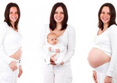
Physiological postpartum period: changes in the body of the postpartum woman
Last reviewed: 23.04.2024

All iLive content is medically reviewed or fact checked to ensure as much factual accuracy as possible.
We have strict sourcing guidelines and only link to reputable media sites, academic research institutions and, whenever possible, medically peer reviewed studies. Note that the numbers in parentheses ([1], [2], etc.) are clickable links to these studies.
If you feel that any of our content is inaccurate, out-of-date, or otherwise questionable, please select it and press Ctrl + Enter.
The puerperal, or postpartum period is the period beginning after the birth of the afterbirth and lasting 8 weeks. During this time, reverse development (involution) of organs and systems, which underwent changes due to pregnancy and childbirth, occurs. Exceptions are the mammary gland and hormonal system, the function of which reaches within the first few days of the postpartum period of its maximum development and continues the entire lactation period.
Early and late postpartum period
The early postpartum period begins with the birth of the afterbirth and lasts for 24 hours. This is an extremely important period of time during which important physiological adaptations of the mother's organism to new conditions of existence take place, especially the first 2 hours after childbirth.
In the early postpartum period, there is a threat of bleeding due to violations of hemostasis in the vessels of the placenta site, a violation of contractile activity of the uterus and traumatization of soft birth canal.
The first 2 hours after childbirth the puerperium remains in the delivery room. The obstetrician closely follows the general condition of the woman, her pulse, measures blood pressure, body temperature, constantly monitors the condition of the uterus: determines its consistency, the height of the standing of the uterus in relation to the pubis and the navel, monitors the degree of hemorrhage,
Late postpartum period - comes in 24 hours after birth and lasts 6 weeks.
 [
[Uterus
The most pronounced process of reverse development is observed in the uterus. Immediately after birth, the uterus contracts, acquires a globular shape, 7 a dense consistency. Its bottom is 15-16 cm above the pubis. The thickness of the walls of the uterus, the largest in the region of the bottom (4-5 cm), gradually decreases towards the neck, where the thickness of the muscles is only 0.5 cm. There is a small amount of blood clots in the uterine cavity. The transverse size of the uterus is 12-13 cm, the length of the cavity from the external throat to the bottom is 15-18 cm, weight - about 1000 g. The cervix is freely passable for the hand. Due to the rapid decrease in the volume of the uterus, the walls of the cavity have a folded character, and are subsequently gradually smoothed out. The most pronounced changes in the uterine wall are noted at the location of the placenta - in the placental area, which is a wound rough surface with blood clots in the vascular region. At other sites, parts of the decidua, the remains of the glands are determined, from which the endometrium is subsequently restored. Periodic contractile movements of the uterine musculature are predominantly preserved in the region of the bottom.
During the following week, due to involution of the uterus, its mass decreases to 500 g, by the end of the second week - to 350 g, the third - to 200-250 g. By the end of the postpartum period, it weighs, as in a state outside of pregnancy, - 50-60 g.
The weight of the uterus in the postpartum period decreases due to the constant tonic reduction of muscle fibers, which leads to a decrease in blood supply and, as a consequence, to hypotrophy and even atrophy of individual fibers. Most of the vessels are obliterated.
During the first 10 days after birth, the bottom of the uterus descends daily approximately by one transverse finger (1.5-2 cm) and on the 10th day is at the level of the womb.
Involution of the cervix has some features and is somewhat slower than the body. Changes begin with an internal throat: already 10-12 hours after birth, the inner pharynx begins to contract, decreasing to 5-6 cm in diameter.
External sinus due to a thin muscular wall remains almost the same. The cervical canal in this regard has a funnel-like shape. In a day the channel narrows. By the 10th day, the inner pharynx is practically closed. Formation of the external throat is slower, therefore finally the cervix is formed by the end of the 13th week of the postpartum period. The original shape of the external throat is not restored due to the overgrowing and tearing in the lateral parts during labor. Uterus is a transverse slit, the cervix is cylindrical, and not conical, as before birth.
Simultaneously with the contraction of the uterus, the uterine lining of the uterus is restored due to the epithelium of the basal layer of the endometrium, the wound surface in the area of the parietal decidual shell ends by the end of the 10th day, with the exception of the placenta, whose healing occurs at the end of the 3rd week. The remains of the decidua and blood clots under the action of proteolytic enzymes melt in the postnatal period from the 4th to the 10th day.
In the deep layers of the inner surface of the uterus, mainly in the subepithelial layer, small-cell infiltration is observed during microscopy, which is formed on the 2nd-4th day after birth in the form of a granulation shaft. This barrier protects against penetration of microorganisms into the wall; in the uterine cavity they are destroyed by the action of proteolytic enzymes of macrophages, biologically active substances, etc. During the involution of the uterus, small-cell infiltration gradually disappears.
The process of regeneration of the endometrium is accompanied by postpartum discharges from the uterus - lochia (from buckwheat lochia - genera). Lochias consist of impurities of blood, leukocytes, blood serum, the remains of the decidua. Therefore, the first 1-3 days after birth are bloody discharge (lochia rubra), on the 4th-7th day the lochia becomes serous-sucronic, have a yellowish-brown color (lochia flava), on the 8th-10th day - without blood , but with a large admixture of leukocytes - a yellowish white color (lochia alba), to which gradually (from the 3rd week) mucus from the cervical canal is mixed. Gradually, the number of lochies decreases, they acquire a mucous character (lochia serosa). On the 3-5th week of excretion from the uterus cease and become the same as before pregnancy.
The total number of licks in the first 8 days of the postpartum period reaches 500-1500 g; they have an alkaline reaction, a specific odor. If, for some reason, lousy lingus retention occurs in the uterine cavity, a lochiometer is formed. In case of infection, an inflammatory process may develop - endometritis.
Fallopian tubes during pregnancy and childbirth due to increased blood filling and edema are thickened and elongated. In the postpartum period, hyperemia and edema gradually disappear. On the 10th day after delivery a complete involution of the fallopian tubes occurs.
In the ovaries in the postpartum period, the regress of the yellow body ends and the maturation of the follicles begins. As a result of the allocation of a large amount of prolactin in nursing women, menstruation is absent for several months or the entire time of breastfeeding. After cessation of lactation, most often after 1.5-2 months, the menstrual function resumes. In some women, ovulation and the onset of pregnancy are possible during the first months after childbirth, even against the background of feeding the baby.
In most non-breasted women, menstruation resumes at 6-8 weeks after delivery.
The vagina after delivery is widely disclosed. The lower sections of its walls protrude into the gaping genital slit. The walls of the vagina are swollen, blue-purple in color. On their surface, cracks and abrasions are revealed. The lumen of the vagina in the primiparous, as a rule, does not return to its original state, but remains broader; folds on the walls of the vagina are less pronounced. In the first weeks of the postpartum period, the volume of the vagina is reduced. Abrasions and tears heal by the 7-8th day of the postpartum period. From the hymen remain papillae (carunculae myrtiformis). The genital gash closes, but not completely.
The ligamentous apparatus of the uterus is restored mainly by the end of the 3rd week after childbirth.
Muscles of the perineum, if they are not injured, begin to restore their function in the first days and acquire an ordinary tone to the 10th-12th day of the postpartum period, the muscles of the anterior abdominal wall gradually restore their tone to the 6th week of the postpartum period.
Mammary gland
The function of the mammary glands after childbirth reaches its highest development. During pregnancy, milk ducts form under the action of estrogens, proliferation of glandular tissue occurs under the influence of progesterone, prolactin-enhanced blood flow to the mammary glands and their engorgement, most pronounced on the 3rd-4th day of the postpartum period.
In the postpartum period, the following processes occur in the mammary glands:
- mammogenesis - development of the breast;
- lactogenesis - initiation of milk secretion;
- galactopoiesis - maintenance of milk secretion;
- galactokinesis - removal of milk from the gland,
The secretion of milk occurs as a result of complex reflex and hormonal effects. The formation of milk is regulated by the nervous system and prolactin. Stimulant action has hormones of the thyroid gland and adrenal glands, as well as reflex action in the act of sucking,
Blood flow in the mammary gland increases significantly during pregnancy and later during lactation. There is a close correlation between blood flow velocity and milk secretion rate. The milk accumulated in the alveoli can not passively enter the ducts. This requires a reduction in the surrounding ducts of myoepithelial cells. They cut the alveoli and push the milk into the duct system, which contributes to its isolation. Myoepithelial cells, like myometrium cells, have specific receptors for oxytocin.
Adequate isolation of milk is an important factor in successful lactation. First, while alveolar milk is available to the child and, secondly, the removal of milk from the alveoli is necessary to continue the day of its secretion. Therefore, frequent feeding and emptying the breast improves milk production.
The increase in the production of milk is usually achieved by increasing the frequency of feeding, including feeding at night, and in the case of insufficient sucking activity in the newborn - feeding one by one, then another by the mammary gland. After cessation of lactation, the mammary gland usually assumes its original dimensions, although the glandular tissue does not completely regress.
Ingredients of breast milk
The secret of the mammary glands secreting in the first 2-3 days after delivery is called colostrum, a secret that is released on the 3-4th day of lactation - transitional milk, which gradually turns into ripe breast milk.
Colostrum
Its color depends on the carotenoids included in the colostrum. Relative density of colostrum 1,034; dense substances account for 12.8%. The composition of colostrum includes colostrum, leukocytes and milk balls. Colostrum is richer than mature breast milk with proteins, fats and minerals, but is poorer in carbohydrates. The energy value of the colostrum is very high: on the first day of lactation it is 150 kcal / 100 ml, on the 2nd - 110 kcal / 100 ml, in the third - 80 kcal / 100 ml.
The amino acid composition of colostrum occupies an intermediate position between the amino acid composition of breast milk and blood plasma.
The total content of colostrum immunoglobulins (which are mainly antibodies) in classes A, C, M and O exceeds their concentration in breast milk, so that it actively protects the newborn's body.
The colostrum also contains a large amount of oleic and linoleic acids, phospholipids, cholesterol, triglycerides, which are necessary structural elements of cell membranes, myelinated nerve fibers, etc. Carbohydrates besides glucose include sucrose, maltose and lactose. On the second day of lactation, the greatest amount of beta-lactose was observed, which stimulates the growth of bifidobacteria that prevent the proliferation of pathogenic microorganisms in the intestine. In colostrum, a large number also contains mineral substances, vitamins, enzymes, hormones and prostaglandins.
Breast milk is the best kind of food for a child of the first year of life. The amount and ratio of the main ingredients that make up the milk of women provide the optimal conditions for their digestion and absorption in the digestive tract of the child. The difference between female and cow's milk (which is most often used for feeding a child in the absence of breast milk) is very significant.
Proteins of female milk are ideal, their biological value is 100%. Breast milk contains protein fractions identical to serum. In breast milk proteins, there is much more albumin, while in cow's milk there is more caseinogen.
Mammary glands are also part of the immune system, specifically adapted to provide the immune protection of the newborn from infections of the digestive and respiratory tract.
The cardiovascular system
After delivery, the bcc decreases by 13.1%, the volume of circulating plasma (CGT) - by 13%, the volume of circulating red blood cells - by 13.6%.
The decrease in BCC in the early postpartum period is 2-2.5 times higher than the blood loss and is due to the deposition of blood in the abdominal organs with a decrease in intra-abdominal pressure immediately after childbirth.
Further, BCC and CGT increase due to the transition of extracellular fluid into the vascular bed.
OCS and circulating hemoglobin content remain reduced throughout the postpartum period.
Heart rate, stroke volume and cardiac output immediately after childbirth remain elevated and in some cases higher for 30-60 minutes. During the first week of the postpartum period, the baseline values of these indicators are determined. Up to the 4th day of the postpartum period, a transient increase in systolic and diastolic pressure may be observed by approximately 5%
The urinary system
Immediately after birth, hypotension of the bladder and a decrease in its capacity are observed. Hypotension of the bladder is aggravated by prolonged labor and the use of ziduralnoy anesthesia. Hypotension of the bladder causes difficulty and violation of urination. The parlor can not feel the urge to urinate or they become painful.
Organs of digestion
Due to some atony of the smooth muscles of the digestive tract, constipation can be observed, which disappear with a rational diet and an active lifestyle. Hemorrhoids often appear after birth (if they are not infringed), little worries about the puerperas.